Tracking alopecia areata accurately is the only way to know whether your treatment is working, your disease is progressing, or your condition is stable. Misdiagnosis of hair loss type leads to wrong treatment in 28% of cases, so starting with an accurate assessment of what you are dealing with is the foundation. This guide covers exactly how to monitor your alopecia areata using methods that produce reliable, comparable data over time.
This article is for informational purposes only and does not constitute medical advice.
Why Tracking Matters for Alopecia Areata
Alopecia areata is unpredictable. Patches can expand, shrink, stay the same, or multiply over weeks to months. Without consistent tracking, it is nearly impossible to determine whether changes are treatment-related or part of the natural disease course. Your dermatologist needs objective data to make treatment decisions, and subjective impressions like "I think it's getting worse" are not enough.
Good tracking also helps you:
- Identify triggers by correlating flares with life events, illness, or stress periods
- Document treatment response for insurance purposes if you need to escalate to systemic therapy
- Catch new patches early when they are most responsive to treatment
- Reduce anxiety by replacing uncertainty with data
Step 1: Establish a Photo Protocol
Photography is the backbone of alopecia areata tracking. The key is consistency across every session.
Equipment
You do not need professional equipment. A smartphone camera works well as long as you follow these rules:
- Same device: Use the same phone or camera each time
- Same distance: Mark a position for your camera or hold it at a fixed distance (arm's length for selfies, or use a tripod/phone mount)
- Same lighting: Natural daylight from a window is best. Overhead bathroom lights create shadows that change the appearance of patches. Photograph at the same time of day
- Same angles: Shoot from at least four positions: front, left side, right side, and top (crown). For patches in specific locations, add close-up shots of each patch
Frequency
- During active treatment: Weekly photos of all affected areas
- During stable periods: Biweekly or monthly photos
- When you notice changes: Photograph immediately and note the date
Organization
Create a dedicated album or folder labeled with the month and year. Name photos with the date and view angle (for example, "2026-02-23-left-side"). This makes it simple to compare images from different sessions side by side.
Step 2: Measure Patch Size
Visual comparison alone can be misleading. Adding measurements creates objective data points.
Simple Measurement Method
Use a small, flexible measuring tape or a ruler placed next to each patch. Measure the longest diameter and the perpendicular diameter of each patch. Record both numbers.
| Patch | Location | Date | Length (cm) | Width (cm) | Notes |
|---|---|---|---|---|---|
| Patch 1 | Left temporal | 2026-01-15 | 3.2 | 2.8 | Active edges |
| Patch 1 | Left temporal | 2026-02-15 | 3.0 | 2.5 | Vellus hairs at edges |
| Patch 2 | Crown | 2026-01-15 | 1.5 | 1.5 | New patch |
| Patch 2 | Crown | 2026-02-15 | 1.5 | 1.4 | Stable |
SALT Score
The Severity of Alopecia Tool (SALT) score is the clinical standard for measuring alopecia areata extent. It divides the scalp into four quadrants and estimates the percentage of hair loss in each:
- Top of scalp (vertex): 40% of total scalp area
- Right side: 18% of total scalp area
- Left side: 18% of total scalp area
- Back (posterior): 24% of total scalp area
Your total SALT score is the sum of the percentage of hair loss in each quadrant multiplied by that quadrant's weight. A SALT score of 0 means no hair loss; 100 means complete scalp hair loss.
You do not need to calculate this precisely at home, but understanding the system helps you communicate with your dermatologist. Even a rough estimate like "my SALT score has gone from around 15 to about 10 since starting treatment" is useful clinical information.
Step 3: Track Symptoms and Triggers
Alongside visual tracking, maintain a simple log of factors that may influence your disease:
- Stress levels: Rate daily stress on a 1 to 10 scale
- Sleep quality: Hours slept and subjective quality
- Illness: Any infections, colds, or other health events
- Medications: Any changes to prescriptions or supplements
- Diet changes: Significant dietary shifts
- Hormonal events: Menstrual cycle, pregnancy, menopause
- Treatment adherence: Whether you applied/took prescribed treatments as directed
Over months, patterns may emerge. Many patients discover that their flares follow specific triggers by 2 to 6 weeks. For a detailed understanding of how these triggers work, read about alopecia areata causes and triggers.
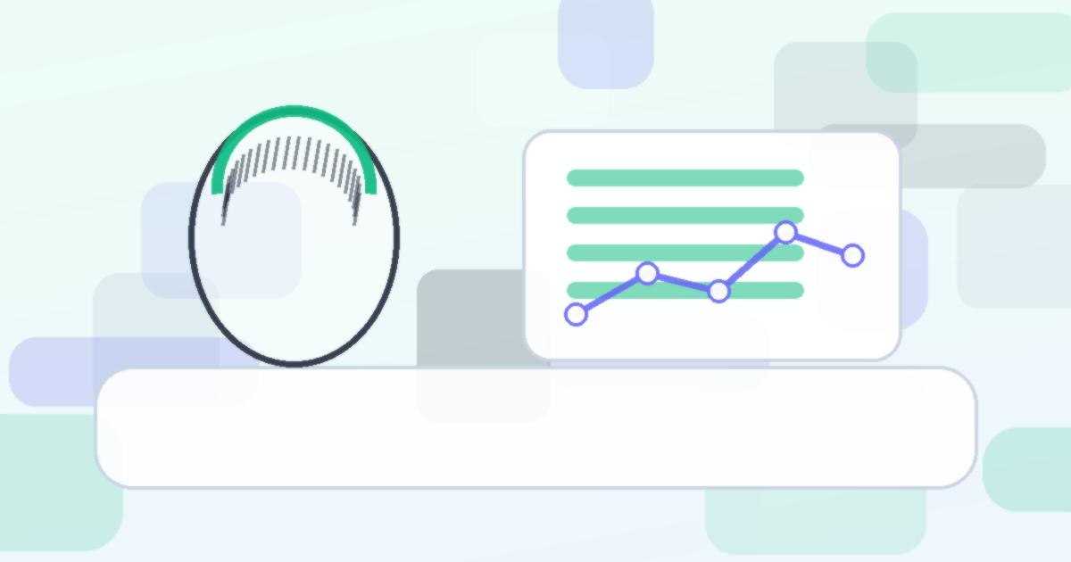
Step 4: Use AI-Assisted Monitoring
AI-powered assessment tools can provide an additional layer of tracking by analyzing photos with consistency that human observation sometimes lacks. These tools are particularly useful for detecting subtle changes in hair density at patch margins, quantifying vellus hair regrowth that may not be obvious to the naked eye, and providing standardized comparisons between photo sessions.
AI monitoring works best as a supplement to dermatologist visits, not a replacement. The combination of professional clinical assessment and consistent at-home tracking gives you the most complete picture.
Step 5: Prepare for Dermatology Appointments
Your tracking data becomes valuable when you bring it to your dermatologist appointments. Before each visit:
- Organize photos: Select comparison shots showing the same patches at different dates
- Summarize measurements: Bring your patch size log showing trends
- Note questions: Write down anything you noticed in your tracking that you want to discuss
- Treatment diary: Record any side effects or compliance issues
Arrive with this prepared, and your appointment becomes far more productive. Your dermatologist can make better treatment decisions with objective trend data rather than relying on a snapshot from a single visit.
Step 6: Know What Regrowth Looks Like
Understanding the stages of regrowth helps you interpret what you are seeing:
- Vellus hair: Fine, colorless peach fuzz. This is the earliest sign of regrowth and a positive signal
- Intermediate hairs: Slightly thicker and may have some pigment. Still finer than normal terminal hair
- Terminal hair: Full-thickness, pigmented hair. This indicates mature regrowth
- White regrowth: Hair that returns without pigment initially. This is common in alopecia areata and often darkens over months
Seeing vellus hairs in a previously bald patch means follicles are reactivating. This is progress, even though the patch may not yet look "filled in."
Build Your Tracking Habit
Start with what is manageable. Even just taking consistent weekly photos from the same angles under the same lighting puts you ahead of most patients. As the habit forms, add measurements and the symptom log. The data you collect gives both you and your medical team the information needed to make the best treatment decisions.
For those considering whether future surgical options might be appropriate after disease stabilization, see the hair transplant candidacy assessment.
Get your free AI hair analysis at myhairline.ai/analyze