
TL;DR: A scalp biopsy removes one or two small skin samples from your scalp under local anesthetic and sends them to a dermatopathologist. It's the most reliable way to tell apart androgenetic alopecia, alopecia areata, scarring alopecias, and telogen effluvium when a clinical exam leaves the diagnosis uncertain. Results typically take one to two weeks and directly determine which treatments are appropriate.
What is a scalp biopsy and why does it matter for female hair loss?
A scalp biopsy is a minor in-office procedure where a dermatologist removes a small core of scalp tissue, usually with a 4mm punch tool, and sends it to a dermatopathologist for microscopic analysis. The sample captures the full hair follicle architecture, from the surface epidermis down to the subcutaneous fat where the deepest follicle bulb sits.
For women, getting the diagnosis right matters more than most people realize. The four most common causes of female hair loss look nearly identical in a mirror: thinning at the part line, reduced density at the crown, a visible scalp through the hair. But their underlying biology is completely different, and treatments that help one condition can actively worsen another. Finasteride, which works well for androgenetic alopecia, has no proven role in scarring alopecias. High-dose corticosteroids are the backbone of alopecia areata therapy but do nothing for androgenetic alopecia. Misdiagnosis means months or years of lost time and money.
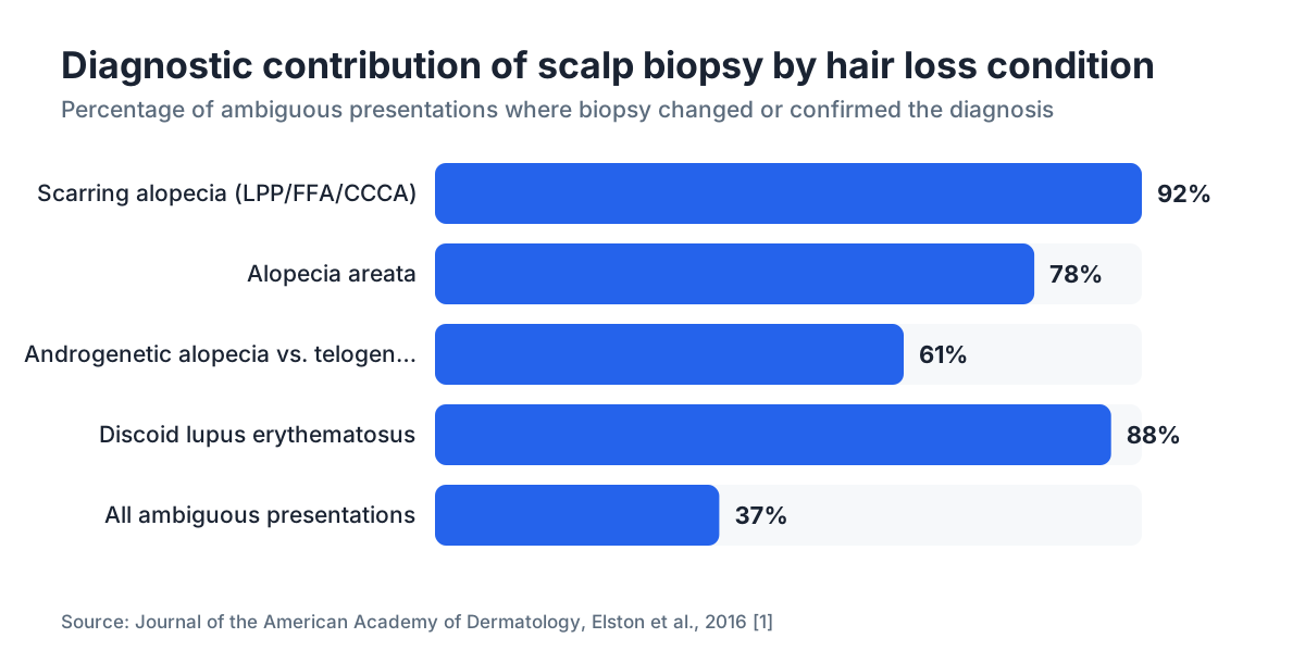
A 2016 study published in the Journal of the American Academy of Dermatology found that scalp biopsy changed or refined the clinical diagnosis in approximately 37% of cases where the presenting picture was ambiguous [1]. That's not a small number. Roughly one in three women who gets a biopsy in this context learns something the exam alone missed.
The procedure is not always necessary. If the pattern is textbook and bloodwork rules out thyroid disease, iron deficiency, and autoimmune markers, a dermatologist may be comfortable treating empirically. But ambiguous presentations, rapid progression, or suspicion of a scarring alopecia are clear indications to biopsy before committing to any treatment.
When do doctors recommend a scalp biopsy?
Not every woman with thinning hair needs a biopsy. Dermatologists generally recommend one when at least one of the following is true.
The clinical picture is genuinely unclear. If a woman has diffuse shedding that could be telogen effluvium or could be early androgenetic alopecia, bloodwork and the Ludwig scale alone often can't separate them reliably. Biopsy can.
Scarring alopecia is in the differential. Conditions like lichen planopilaris (LPP), frontal fibrosing alopecia (FFA), and central centrifugal cicatricial alopecia (CCCA) destroy follicles permanently. The window to halt progression is narrow. A clinical exam can suggest these diagnoses but cannot confirm them without histology. The American Academy of Dermatology guidelines on hair loss strongly support biopsy when scarring alopecia is suspected [2].
The patient has symptoms that don't fit a standard pattern. Tingling scalp and female hair loss is a pairing worth taking seriously. Perifollicular tingling, burning, or tenderness often signals active inflammation, which is a hallmark of lichen planopilaris and FFA. When a woman describes a crawling or burning sensation at her hairline alongside recession, biopsy is more urgent, not less, because those symptoms suggest active scarring is underway.
Treatment has failed. If a woman has been on minoxidil for twelve months with zero response, confirming or ruling out androgenetic alopecia before escalating is reasonable.
There is patchy or irregular loss. Patchy loss could be alopecia areata, traction alopecia, secondary syphilis, or tinea capitis. Only histology, sometimes combined with culture, can reliably separate these.
What conditions can a scalp biopsy diagnose?
The table below summarizes the main conditions a dermatopathologist looks for in female hair loss biopsies, their key histological findings, and whether the hair loss is reversible.
| Condition | Key biopsy findings | Reversibility |
|---|---|---|
| Androgenetic alopecia (FPHL) | Miniaturized follicles, increased vellus:terminal ratio >2.5:1, mild perifollicular fibrosis | Partial with treatment |
| Telogen effluvium | Elevated telogen follicle count (>15-20%), normal follicle size | Usually full |
| Alopecia areata | Lymphocytic infiltrate around anagen bulbs ("swarm of bees"), nanogen follicles | Variable |
| Lichen planopilaris | Lichenoid infiltrate, follicular destruction, perifollicular fibrosis, loss of sebaceous glands | No (scarring) |
| Frontal fibrosing alopecia | LPP-like changes at frontal hairline, vellus follicle involvement | No (scarring) |
| CCCA | Premature desquamation of inner root sheath, lamellar fibrosis | No (scarring) |
| Discoid lupus | Interface dermatitis, mucin deposition, pigment incontinence | Partial if caught early |
| Tinea capitis | Fungal hyphae within hair shaft (PAS stain) | Yes with antifungals |
Androgenetic alopecia, also called female pattern hair loss (FPHL), is the most common diagnosis in premenopausal and postmenopausal women. Its hallmark on biopsy is miniaturization: follicles that have progressively shrunk from terminal (thick, pigmented) to vellus (thin, unpigmented). A vellus-to-terminal ratio above 2.5:1 is considered diagnostic [3].
Telogen effluvium shows a different picture: the follicles themselves are normal size, but an abnormally high percentage are resting rather than actively growing. This is what happens after major physical stress, crash dieting, postpartum hormone shifts, or thyroid dysfunction. The follicles are intact. They just need the trigger removed.
The scarring alopecias (LPP, FFA, CCCA, discoid lupus) are the conditions where biopsy is most urgently needed, because they are irreversible once the follicle is replaced by scar tissue. Early biopsy is the difference between halting the disease and watching permanent loss compound.
How is a scalp biopsy performed, step by step?
The whole thing takes about fifteen minutes in a dermatologist's office. No hospital. No general anesthesia.
First, the dermatologist selects the biopsy site. For diffuse hair loss, they typically target an actively affected area, often at the vertex or mid-scalp, avoiding completely bald spots where follicles are already gone. For suspected scarring alopecias, they aim at the active edge of the lesion, where inflammation is still happening, because a burned-out scar yields no useful information.
Some clinicians take two samples: one processed horizontally (transverse sections) and one vertically. Horizontal sectioning is now widely preferred for hair loss workups because a single 4mm punch yields cross-sections of 20-30 follicles, giving the pathologist far more statistical power than the 4-5 follicles visible on a vertical section [4].
The site is numbed with a small injection of lidocaine. Most people describe this as a brief sting and then nothing. The punch biopsy tool is pressed into the scalp, rotated, and the core lifted out with forceps and scissors. The resulting hole is typically closed with one or two sutures, or sometimes just a pressure dressing if very small.
The samples go to the lab in formalin fixative. Specialized stains are used depending on clinical suspicion: hematoxylin and eosin (H&E) for standard morphology, periodic acid-Schiff (PAS) for fungal elements, direct immunofluorescence (DIF) if lupus is suspected.
Sutures come out in 7-14 days. Scarring is minimal and usually hidden by surrounding hair.
Does a scalp biopsy hurt?
Most patients say the lidocaine injection is the worst part, a quick pinch and burn that fades within seconds. The punch itself is painless. After the anesthetic wears off, the area is tender for a day or two, and over-the-counter acetaminophen or ibuprofen handles it easily.
The scalp is a highly vascular structure, so there is often a small amount of bleeding during the procedure. This is normal and controlled with pressure. Infection is possible but uncommon; the dermatologist will give specific wound care instructions.
The small scar that remains is typically invisible once the surrounding hair grows back. Most women report the procedure is significantly less uncomfortable than they anticipated.
What does a scalp biopsy cost, and does insurance cover it?
Out-of-pocket costs vary widely. A single punch biopsy procedure in the United States typically ranges from $150 to $400 for the office visit and biopsy itself. Dermatopathology laboratory fees add another $100 to $300 or more, billed separately by the lab [5].
Most insurance plans, including Medicare, cover scalp biopsy when it is medically indicated and the claim is coded appropriately. CPT code 11305 covers shave biopsy of the scalp; 11104 covers punch biopsy. The key word is "medically indicated." If the dermatologist documents clinical ambiguity or suspicion of a serious condition, coverage is usually approved. Cosmetic hair loss alone, coded as such, may be denied.
Prior authorization is sometimes required. Call your insurer before the appointment, confirm coverage under your specific plan, and ask the dermatologist's office to submit the correct ICD-10 code for the suspected diagnosis rather than just "hair loss, unspecified" (L65.9), which insurers sometimes interpret as cosmetic.
If you are uninsured, asking for the cash-pay rate before the appointment often yields a meaningfully lower price than the billed rate.
How accurate is a scalp biopsy compared to other diagnostic tests?
No single hair loss diagnostic is perfect, but biopsy comes closest.
Bloodwork (thyroid panel, ferritin, complete blood count, ANA, DHEA-S, free testosterone) rules out systemic contributors but cannot tell you what is happening at the follicle level. A woman can have normal labs and active LPP simultaneously.
Dermoscopy (trichoscopy) is a noninvasive technique where the dermatologist examines the scalp under magnification. In experienced hands it can strongly suggest specific diagnoses. A 2013 review in the Journal of Investigative Dermatology Symposium Proceedings noted trichoscopy has high sensitivity for conditions like alopecia areata (yellow dots, exclamation-mark hairs) and LPP (perifollicular scaling, loss of follicular openings) [6]. It's a useful screening step. But trichoscopy is operator-dependent and cannot confirm early histologic changes that precede visible surface signs.
Pull test and tug test are quick clinical assessments but have poor specificity. A positive pull test tells you follicles are shedding; it says nothing about why.
Biopsy is the only test that directly examines follicle structure, the ratio of follicle sizes, the presence and type of inflammatory infiltrate, and the degree of fibrosis. For suspected scarring alopecia, it is not a tiebreaker, it's the required standard of care.
The limitation of biopsy is sampling error. A 4mm punch samples a tiny area of the scalp. If the active disease is patchy and the biopsy misses the active edge, the result can be falsely reassuring. Experienced dermatologists minimize this by choosing the biopsy site carefully.
How do biopsy results change your treatment plan?
This is the whole point of the procedure. The results directly determine which treatments are appropriate and which ones to avoid.
If the biopsy confirms androgenetic alopecia (FPHL), the evidence-based options are minoxidil topical or oral minoxidil, spironolactone, and in some countries low-dose finasteride off-label. You can also look at dht blockers and, if loss is advanced, consider whether hair transplant candidacy applies. Knowing the diagnosis is FPHL and not something else means you can commit to a multi-year treatment plan with confidence.
If the biopsy shows telogen effluvium, the first move is removing the trigger if one is identifiable. Heavy shedding after stopping or starting medications, after surgery, after a high-stress period, or postpartum usually resolves on its own within 6-12 months. Minoxidil can help support regrowth. Minoxidil side effects are worth reviewing before starting, especially for women considering the oral route.
If the biopsy confirms a scarring alopecia like LPP or FFA, the entire treatment approach shifts. The priority is stopping inflammation before more follicles are destroyed, not regrowing hair. First-line options include topical and intralesional corticosteroids, hydroxychloroquine, and, in some cases, oral retinoids or JAK inhibitors. Using minoxidil as a primary treatment here is not wrong, but it does nothing to address the underlying inflammatory process.
If the biopsy suggests alopecia areata, treatment options include intralesional triamcinolone injections, topical or systemic immunosuppressants, and, since 2022, FDA-approved JAK inhibitors (baricitinib received FDA approval for severe alopecia areata in June 2022 [7]).
Knowing the exact diagnosis also tells you what not to spend money on. Women who screen themselves at home using tools like the free AI hair analysis at MyHairline before a dermatologist visit can bring more structured questions to that appointment, which often helps the clinician decide faster whether a biopsy is warranted.
What is the connection between a tingling scalp and female hair loss?
Tingling, burning, itching, or tenderness on the scalp alongside hair thinning is not something to dismiss. It is a clinically meaningful symptom.
In the context of frontal fibrosing alopecia and lichen planopilaris, perifollicular burning or tenderness is a marker of active inflammation. A 2019 study in JAMA Dermatology found that patients with FFA frequently report scalp pruritus and tenderness, and that symptom presence correlated with disease activity [8]. The practical implication: if you have a receding frontal hairline combined with tingling or burning at the scalp, treat this as urgent and see a dermatologist promptly. Delay means permanent follicle loss.
Tingling can also stem from contact dermatitis (a reaction to a shampoo, hair dye, or topical minoxidil itself), seborrheic dermatitis, or psoriasis. These are not hair-loss conditions per se, but scalp inflammation can contribute to shedding and can make pattern loss look worse. Biopsy is usually not the first step here; a clinical exam often suffices.
Less commonly, scalp tingling with hair loss points toward discoid lupus erythematosus, which requires DIF on biopsy to confirm. This matters because lupus-related hair loss can be scarring or non-scarring depending on the type, and systemic workup may follow.
Here's the takeaway: tingling scalp plus hair loss is a reason to escalate beyond basic bloodwork, not a reason to reassure yourself with a moisturizing shampoo.
Are there risks or reasons to avoid a scalp biopsy?
The procedure is low-risk. Serious complications are rare. The realistic risks are a small scar (usually hidden by hair), temporary tenderness, a very small chance of infection, and the slight theoretical risk that a suture causes a cyst.
Allergy to lidocaine is uncommon but possible. Patients should disclose all allergies before the procedure.
Blood thinners (warfarin, apixaban, aspirin) increase bleeding risk. Most dermatologists ask patients to hold aspirin and NSAIDs for a few days before the procedure if clinically safe, and to discuss anticoagulant management with their prescribing physician.
Biopsy is not useful in truly burned-out, end-stage scarring alopecia where only fibrotic tissue remains. In that scenario, the histology shows nonspecific fibrosis and gives no actionable information. The time to biopsy is before that stage.
Pregnancy is not a contraindication, but the dermatologist will select lidocaine formulations without epinephrine in some cases and document the clinical necessity. The AAD does not list pregnancy as a contraindication to diagnostic skin biopsy [2].
The risk-to-benefit math strongly favors biopsy when the clinical indication is present. The information gained almost always outweighs the minor procedural discomfort.
How should you prepare for a scalp biopsy appointment?
Bring a written list of all medications and supplements. Some supplements, particularly fish oil and vitamin E at high doses, have mild antiplatelet effects and may be worth pausing if your dermatologist advises it.
Wash your hair the morning of the appointment but do not apply styling products. The dermatologist needs a clear view of the scalp.
Bring photos of your hair from 6-12 months ago if you have them. Progression pattern and speed matter when interpreting results, and side-by-side photos are genuinely useful clinical information.
Ask in advance whether direct immunofluorescence will be performed. DIF requires the sample to be sent fresh (in Michel's transport medium), not formalin-fixed. If lupus is in the differential and you don't ask, the lab might receive the wrong sample type and the DIF result will be uninterpretable.
Arrange to have someone drive you if you are anxious about the procedure, though most people drive themselves home without any issue. The anesthetic resolves within an hour, and the wound is dressed before you leave.
After the procedure, keep the area dry for 24-48 hours, apply any antibiotic ointment as directed, and return to have sutures removed on the schedule given. Hair washing can usually resume gently after 48 hours.
What happens after you get your scalp biopsy results?
Results typically return in 7-14 business days, sometimes faster if the lab is in-house. Your dermatologist should schedule a follow-up appointment rather than delivering results by a portal message, because the findings usually require explanation and a treatment conversation.
Ask for a copy of the pathology report. You are entitled to it, and having it lets you seek a second opinion if the diagnosis feels uncertain or if you are referred to another specialist.
If the result is non-specific or inconclusive, discuss whether a repeat biopsy at a different site is warranted. An uninformative result from a poor sample site is not the same as a normal result.
Once you have a confirmed diagnosis, you can start building a realistic treatment timeline. Androgenetic alopecia responds slowly to treatment; most clinicians evaluate minoxidil response at 6-12 months. Scarring alopecia requires monitoring every 3-6 months to assess whether inflammation is controlled. What causes hair loss and how the various conditions progress is worth reading as background once you have a diagnosis in hand.
For women whose biopsy confirms androgenetic alopecia and who want to track visible response to treatment over time, the MyHairline free AI scan (/scan) can provide standardized before-and-after scalp analysis to supplement your dermatologist visits.
Sources
- Journal of the American Academy of Dermatology, Elston et al., 2016
- American Academy of Dermatology, Hair Loss Clinical Guidelines
- Whiting DA, Journal of the American Academy of Dermatology, 1993, Diagnostic and predictive value of horizontal sections of scalp biopsy
- Elston DM et al., Journal of Cutaneous Pathology, Horizontal sectioning of the scalp biopsy
- Healthcare Bluebook, Skin Biopsy Cost Data
- Rudnicka L et al., Journal of Investigative Dermatology Symposium Proceedings, 2013, Trichoscopy update 2011
- FDA Drug Approval: Baricitinib (Olumiant) for Alopecia Areata, June 2022
- Vañó-Galván S et al., JAMA Dermatology, 2019, Frontal fibrosing alopecia: multicenter review
- National Institute of Arthritis and Musculoskeletal and Skin Diseases (NIAMS), NIH, Alopecia Areata
- Stefanato CM, Journal of Cutaneous Pathology, 2010, Histopathology of alopecia: a clinicopathological approach to diagnosis
- CMS Medicare, Skin Biopsy CPT Code Coverage