
TL;DR: Trichoscopy is a non-invasive scalp exam where a dermatologist uses a magnifying lens (usually 10x to 70x) to study hair shafts, follicular openings, and scalp skin. In one office visit it can separate androgenetic alopecia from alopecia areata, scarring alopecia, and telogen effluvium, often without a biopsy. Sensitivity for androgenetic alopecia runs about 98 percent.
What is trichoscopy and how does it work?
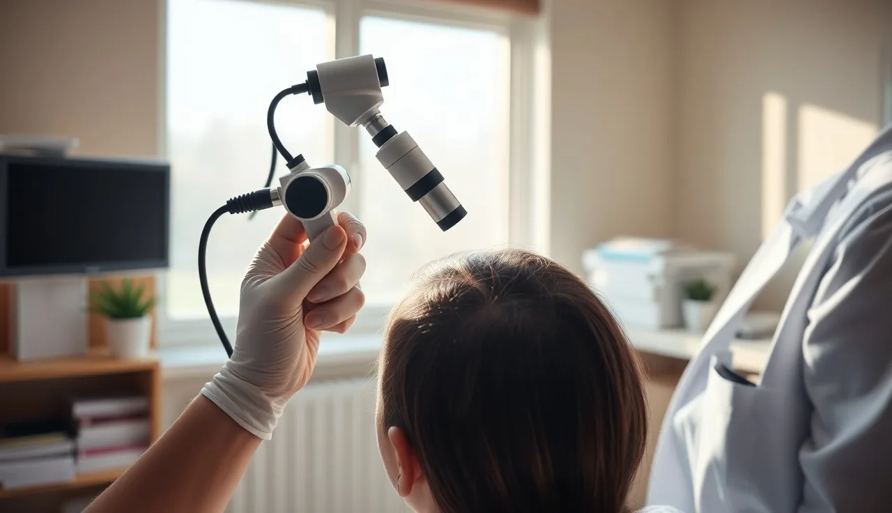
Trichoscopy is dermoscopy pointed at the scalp instead of a mole. A dermatologist presses a dermoscope (a handheld magnifying lens with a polarized light source) against your scalp, or connects it to a digital camera for higher magnification and image storage. Basic handheld devices show 10x. Videodermoscopy systems reach 70x or more. That range is enough to see individual hair shafts, the tiny openings in the scalp called follicular ostia, blood vessel patterns, and pigment deposits the naked eye misses completely.
The exam takes five to fifteen minutes. No needles, no biopsy punch, no local anesthetic. The doctor applies a drop of immersion fluid (or uses polarized light without fluid) and presses the lens against the scalp at several sites. Many clinics photograph each site and store the images to compare progression or treatment response at follow-up.
The technique was formalized in the late 2000s, largely through the work of dermatologist Lidia Rudnicka and colleagues, who published the first systematic trichoscopy criteria. Their 2008 paper in the Journal of the American Academy of Dermatology described the dermoscopic patterns across different alopecia types and became a founding reference for the field [1].
Trichoscopy does not replace a scalp biopsy in every case. Scarring alopecias in particular still often need histopathology to pin down the exact subtype. But for the two most common conditions, androgenetic alopecia and alopecia areata, trichoscopy gives a diagnosis that lines up closely with biopsy findings and spares the patient an invasive procedure.
What structures does a dermatologist actually examine?
A trained eye scans several distinct structures during the exam. Knowing what each one is helps you read the report you might get handed.
Hair shaft diameter. On a healthy scalp, most hairs are roughly the same thickness. Androgenetic alopecia drives progressive miniaturization, meaning follicles shrink and pump out thinner, shorter hairs over time. Dermatologists compare the ratio of thick (terminal) hairs to thin (vellus or miniaturized) hairs in a given patch. A miniaturization rate above 20 percent in the frontal or vertex zone counts as diagnostically significant for androgenetic alopecia [2].
Follicular units. A follicular unit is a cluster of one to four hairs coming out of a single opening. Trichoscopy lets the doctor count how many hairs each unit produces. A healthy scalp shows plenty of two and three hair units. In androgenetic alopecia, single-hair units take over as follicles lose their neighbors.
Follicular openings (ostia). Empty openings, called yellow dots or black dots depending on what fills them, flag different problems. Yellow dots (infundibula plugged with sebum and keratin) are a hallmark of alopecia areata and also show up in androgenetic alopecia. Black dots are broken or cadaverized hairs still stuck inside the follicle, which points toward alopecia areata or trichotillomania [3].
Perifollicular signs. Brown or gray rings around openings (perifollicular discoloration or casts) suggest inflammation or scarring. White dots mark fibrous tracts where follicles have been destroyed for good.
Blood vessel patterns. The dermal blood supply looks different across conditions. Simple honeycomb vessels are normal. Arborizing or twisted red vessels in patchy areas can signal inflammation from lichen planopilaris or discoid lupus.
Scalp surface and interfollicular skin. Scaling, pustules, and pigment changes between openings all feed the diagnosis. Diffuse white scaling favors seborrheic dermatitis or psoriasis as a contributor. Fibrosis between follicles, which looks like a white honeycomb, marks end-stage scarring alopecia.
How does trichoscopy distinguish the most common types of hair loss?
This is where the technique earns its keep. Different alopecia types leave their own fingerprints at the follicle, and a skilled dermatologist can tell them apart fast.
| Condition | Key trichoscopy findings |
|---|---|
| Androgenetic alopecia (AGA) | Hair diameter variability >20%, more single-hair units, peripilar sign (brown halo), yellow dots |
| Alopecia areata | Black dots, yellow dots (empty follicles), exclamation-mark hairs, short vellus hairs in regrowing patches |
| Telogen effluvium | Mostly single-hair follicular units, low density without much miniaturization, upright regrowing hairs |
| Lichen planopilaris (scarring) | Perifollicular scaling, loss of follicular openings, white dots, faded perifollicular redness late-stage |
| Frontal fibrosing alopecia | Lonely hairs (isolated single hairs), perifollicular redness, perifollicular scaling at the hairline |
| Trichotillomania | Broken hairs at different lengths, black dots, coiled hairs, flame hairs, no yellow dots |
| Tinea capitis | Comma hairs, corkscrew hairs, black dots, background scaling |
Androgenetic alopecia is far and away the most common diagnosis. In men and women with AGA, the giveaway is hair shaft diameter variability: the same follicular unit shows a thick hair next to a thin one next to a barely-there vellus hair, all because DHT-driven miniaturization moves at different speeds in different follicles [2]. For the mechanism behind that miniaturization, the what causes hair loss explainer walks through it.
Alopecia areata looks nothing like AGA. The patchy, immune-driven destruction of follicles produces black dots (broken cadaverized hairs), yellow dots (empty infundibula), and the exclamation-mark hair (narrower at the base than the tip, shaped like an exclamation point). Spotting exclamation-mark hairs is essentially confirmatory for alopecia areata without a biopsy [3].
Telogen effluvium is the tricky one. No single dramatic finding announces it. The dermatologist mostly sees diffuse density loss, a shift toward single-hair units, and short upright regrowing hairs standing up from the scalp once shedding starts to resolve. The missing piece, and the useful one, is the absence of miniaturization. That absence separates it from AGA, which matters because the treatment paths split completely.
Scarring alopecias like lichen planopilaris kill the follicle for good. Trichoscopy shows white dots where openings have been replaced by scar tissue, perifollicular scaling, and perifollicular redness during active disease. Those findings push the dermatologist toward a biopsy to nail the inflammatory subtype before treatment starts.
What is hair diameter variability and why does it matter so much?
Hair diameter variability is the single most useful number in trichoscopy for the average person worried about thinning. It tells you how far the miniaturization has gone.
On a normal scalp, at least 80 percent of frontal hairs are terminal hairs above a certain thickness (roughly 0.06 mm in most published criteria). Once androgenetic alopecia sets in, the share of thin, miniaturized hairs climbs. The line most dermatologists draw for diagnostic significance is a miniaturization rate above 20 percent, meaning more than one in five hairs in the examined area is miniaturized [2].
That number has practical teeth. A 25 percent miniaturization rate is early; the follicles are still there and still making some kind of hair. A 60 percent rate means most follicles in that zone are compromised. It shapes both the outlook and whether treatments like finasteride or minoxidil for men are likely to produce visible regrowth or just hold the line.
Digital trichoscopy systems measure hair diameter semi-automatically with image analysis software. Videodermoscopy assigns each visible hair a diameter in micrometers and calculates the percentage falling below the terminal threshold. A subjective visual impression becomes a reproducible number. That is genuinely useful for tracking treatment response across six to twelve months.
Can trichoscopy diagnose scarring alopecia?
Yes, with real caveats. Trichoscopy is good at detecting that scarring exists and at spotting active inflammation. It usually cannot tell you exactly which scarring subtype you have. That still takes a biopsy in most cases.
The common scarring alopecias, lichen planopilaris (LPP), frontal fibrosing alopecia (FFA), central centrifugal cicatricial alopecia (CCCA), and discoid lupus erythematosus (DLE), overlap heavily on trichoscopy. All of them eventually show absent follicular openings plus white dots or patches marking fibrosis. Early on, though, some patterns pull them apart.
LPP and FFA both show perifollicular scaling (a white or silver scale gripping the follicle) and perifollicular redness right at the opening. In FFA, the dermatologist checks the hairline for "lonely hairs," solitary hairs left standing because the follicles around them have been destroyed. FFA also hits eyebrows and eyelashes often, and eyebrow trichoscopy can show the same scaling pattern.
CCCA, which disproportionately affects Black women, tends to show white patches and concentric perifollicular fibrosis at high magnification. A 2019 review in the Journal of the American Academy of Dermatology noted that CCCA trichoscopy findings include perifollicular white or gray halos and loss of follicular openings [4].
The practical bottom line: if trichoscopy turns up any scarring pattern, the dermatologist should still biopsy before starting immunosuppressive treatment. The wrong drug for the wrong subtype can speed up the hair loss.
How accurate is trichoscopy compared to a scalp biopsy?
It depends on the diagnosis, and the numbers are better for some than others.
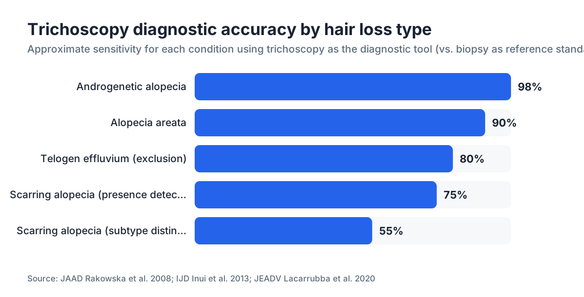
For androgenetic alopecia, trichoscopy performs strongly. A study comparing trichoscopy criteria against scalp biopsy in patients with diffuse hair loss found sensitivity around 98 percent and specificity around 95 percent for AGA when hair diameter variability was the main criterion [2]. Biopsy is not usually needed to confirm uncomplicated AGA.
For alopecia areata, a 2013 study in the International Journal of Dermatology found trichoscopy signs (yellow dots, black dots, exclamation-mark hairs) tracked closely with histopathology, and exclamation-mark hairs were highly specific for the condition [3].
For scarring alopecias, accuracy falls. A 2020 systematic review found trichoscopy correctly flagged the presence of scarring in most cases, but telling LPP from DLE by trichoscopy alone had lower specificity. Biopsy stays the reference standard for scarring subtypes [5].
For telogen effluvium, trichoscopy works by exclusion rather than confirmation. No miniaturization, no yellow dots, no scarring pattern in someone with diffuse shedding supports a TE diagnosis, but the call is ultimately clinical.
Here is the honest summary. Trichoscopy is very accurate for the two most common non-scarring diagnoses (AGA and alopecia areata) and is a genuinely useful first-line, non-invasive tool. For scarring alopecias, treat it as a screening test that guides biopsy decisions, not a replacement for one.
What happens during a trichoscopy appointment?
Most people show up for a hair loss visit and get trichoscopy as part of a standard dermatology exam, not as a separate booking. Here is what to expect.
You will be told not to wash your hair the day of the exam, or to wash it the day before, so some natural sebum is present. Styling products come off first, because they hide follicular openings and create fake findings.
The dermatologist checks multiple scalp regions: the frontal hairline, the vertex (crown), the temporal zones, and the occipital (back) region. The back of the scalp matters because in androgenetic alopecia the occipital zone is generally spared, keeping normal density and no miniaturization. That contrast between a thinning frontal zone and a preserved back is itself a diagnostic clue, and it is exactly why donor hair for hair transplants comes from the occipital region.
The doctor notes findings in each zone and often photographs them. You may see the images on a screen in real time. Some clinics run FotoFinder or similar videodermoscopy platforms that measure hair density (hairs per square centimeter) and mean diameter automatically.
Expect to talk through the findings during the visit. A good dermatologist explains what the images show in plain terms. If you want to track progress between visits, MyHairline's free AI hair scan can capture baseline photos with your phone and flag changes to bring to your dermatologist.
The exam is painless. The only mild discomfort is the lens pressing firmly against the scalp. Total in-room time is usually under twenty minutes.
Does trichoscopy help decide which treatment to use?
Yes, directly. The findings narrow the treatment options a lot.
If the exam confirms androgenetic alopecia with real miniaturization but follicular openings still present, both finasteride and minoxidil for men are on the table. Finasteride cuts DHT, the hormone shrinking the follicles, so it works best when miniaturization is active but not yet finished. A high rate (say, above 50 percent in a zone) suggests those follicles may be too far gone for regrowth, though stabilizing what remains elsewhere is still realistic. For how the two drugs compare when stacked, see finasteride and minoxidil.
If trichoscopy shows alopecia areata, DHT blockers are beside the point. The treatment is immune-directed: topical or intralesional steroids, JAK inhibitors in severe cases, or watchful waiting in mild patchy disease.
If scarring alopecia is on the table, starting a DHT blocker would be a mistake. The job is to shut down the inflammation destroying follicles, usually with hydroxychloroquine or topical tacrolimus for LPP and FFA, not growth-oriented drugs.
For telogen effluvium, no miniaturization on trichoscopy means no permanent loss yet. The focus shifts to finding the trigger (nutritional deficiency, thyroid dysfunction, major stress) rather than starting a hair loss drug. The telogen effluvium article covers those triggers.
Trichoscopy also helps size up whether you are a realistic hair transplant candidate. Dense follicular units in the occipital donor zone mean good graft availability. Miniaturization in the donor zone is a red flag that transplanted hairs may not last.
What do yellow dots, black dots, and white dots each mean?
These three dot patterns show up constantly in trichoscopy reports, and they mean very different things.
Yellow dots are swollen follicular infundibula packed with keratin and sebum. They read as yellow or yellowish-white under the dermoscope. They are strongly tied to alopecia areata (seen in roughly 60 to 70 percent of AA cases) but also turn up in androgenetic alopecia, especially at the vertex. In AA, a yellow dot marks a follicle that shed its hair but stayed structurally intact, which is actually a hopeful sign: regrowth is possible if the immune attack stops.
Black dots are the remains of broken hairs inside the opening, often called cadaverized hairs. They are a hallmark of alopecia areata (in active, spreading lesions) and trichotillomania (where mechanical breakage explains them). In tinea capitis, black dots are fungal-infected hairs snapped off at the scalp surface. Black dots in a patchy bald area make alopecia areata the top diagnosis until proven otherwise [3].
White dots come in two flavors. Small, evenly spaced white dots on normal-looking scalp are just the openings of eccrine sweat glands, a normal finding. Large, irregular white patches or honeycomb-white areas mean fibrotic destruction of follicles, the mark of end-stage scarring alopecia. The distinction is everything: regular white dots mean nothing alarming, white fibrotic patches mean the follicles in that area are gone for good [5].
A single report might mention all three. Reading it right means knowing which scalp zone each dot came from and what else sat alongside it.
How is trichoscopy used to monitor treatment response?
Serial monitoring is one of the most practical uses, and one patients rarely hear about. Trichoscopy images taken three, six, and twelve months apart give an objective record of whether a treatment is working.
Hair loss treatments are slow. Minoxidil needs about three to six months to show visible density changes, and finasteride can take twelve months before most people feel confident it is working. In that waiting period, patients doubt anything is happening at all. Trichoscopy can catch micro-level changes before they show up in the mirror.
A dermatologist monitoring AGA treatment watches for three things: hair diameter climbing (thicker hairs where miniaturized ones were), more multi-hair follicular units, and a falling miniaturization percentage. A 2020 study in Dermatology and Therapy showed patients on oral minoxidil had measurable increases in hair shaft diameter on videodermoscopy within three months, before density improved to the naked eye [6].
For alopecia areata on treatment, the return of regrowing vellus hairs (short, lightly pigmented hairs across a previously bald patch) is the earliest sign of remission. Black dots and yellow dots fade as follicles re-enter the anagen growth phase.
Standardizing the exam location matters. Clinics with digital systems mark reference points on a scalp map so the same 1 cm square is re-examined every visit. Skip that and you are comparing slightly different patches over time, which introduces error.
Tracking your own hair at home works alongside this. Photograph the same spots under consistent lighting each month, then pair that with a dermatologist-read trichoscopy every six months for a fuller picture. The minoxidil side effects article is worth reading if you are starting treatment, because early shedding can look alarming without context.
What equipment does a dermatologist use for trichoscopy?
The basic tool is a dermoscope, a handheld device about the size of an otoscope. Entry-level dermoscopes from brands like Dermlite or Heine cost roughly $400 to $1,200 and offer 10x magnification with polarized light. Most dermatologists already own one for skin cancer screening, so it doubles for scalp work at no extra cost to the practice.
Higher-end videodermoscopy systems, like FotoFinder Trichoscale, Canfield TrichoScan, or Folliscope, bolt a camera to the dermoscope head and show live images on a monitor at up to 70x. These run $5,000 to $50,000 depending on features. They add image storage, automated hair counting, and diameter measurement software. You find them in hair specialist clinics and academic dermatology centers, not most general practices.
TrichoScan deserves a specific mention. It is software that works with standardized dermoscopy images, automates hair density counting (hairs per square centimeter), and splits hairs into anagen and telogen phases based on tip shape. A 2009 validation study found good reproducibility for density measurements, though it needed careful standardization of image capture to stay reliable [7].
Do not be discouraged if your dermatologist uses a basic handheld dermoscope instead of a fancy system. For the questions that matter most (AGA versus AA versus scarring), a trained eye with a $500 dermoscope gives clinically useful answers. The expensive digital gear mostly adds value for serial monitoring and research, not the first diagnosis.
Is trichoscopy covered by insurance and what does it cost?
In the United States, trichoscopy has no CPT billing code of its own. It gets billed inside the dermatology consultation, not as a separate procedure. So if your visit is covered by health insurance, the trichoscopy done during that visit is typically covered as part of the exam. You pay whatever your plan requires for a specialist visit.
In practices that charge separately for scalp analysis (common in hair restoration clinics, less so in medical dermatology offices), a dedicated trichoscopy with digital imaging typically runs $100 to $300 out of pocket based on prices clinics have listed publicly. Pricing varies widely by region and clinic type. There is no standard national price, and these numbers move.
A scalp biopsy, if the findings warrant one, adds cost. A punch biopsy with pathology processing typically runs $200 to $600 or more out of pocket depending on insurance and lab. Trichoscopy's real economic value for patients is that it often avoids the biopsy entirely for straightforward AGA and AA.
For a preliminary read on your pattern before booking a dermatology visit, the free AI hair scan at MyHairline (/scan) gives you a starting point to bring to a doctor. It does not replace clinical trichoscopy.
Sources
- Journal of the American Academy of Dermatology, Rudnicka et al. 2008: Trichoscopy overview
- Journal of the American Academy of Dermatology, Rakowska et al. 2008: Trichoscopy criteria for AGA
- International Journal of Dermatology, Inui et al. 2013: Trichoscopy findings in alopecia areata
- Journal of the American Academy of Dermatology, Callender et al. 2019: CCCA trichoscopy review
- Journal of the European Academy of Dermatology and Venereology, Lacarrubba et al. 2020: Trichoscopy accuracy in scarring alopecia systematic review
- Dermatology and Therapy, Vañó-Galván et al. 2020: Oral minoxidil and trichoscopy response
- British Journal of Dermatology, Hoffmann 2009: TrichoScan validation
- American Academy of Dermatology: Hair loss overview and diagnosis guidance
- National Institutes of Health MedlinePlus: Alopecia areata
- FDA: Minoxidil labeling (OTC topical)
- FDA: Finasteride (Propecia) prescribing information