
TL;DR: Lupus causes hair loss through scalp inflammation (discoid lupus) or a systemic stress response (lupus-related telogen effluvium). Androgenetic alopecia follows a predictable genetic thinning pattern driven by DHT. You tell them apart by loss pattern, scalp appearance, biopsy findings, and blood tests. Getting the diagnosis right matters because the treatments have nothing in common.
Why does lupus cause hair loss in the first place?
Lupus is an autoimmune disease in which the immune system attacks healthy tissue. The scalp and hair follicles can become targets in two distinct ways.
The first is discoid lupus erythematosus (DLE). In DLE, chronic inflammation attacks follicles directly, destroying them and leaving behind scarred patches. Because the follicle itself is destroyed, hair loss in those areas is usually permanent [1]. The American Academy of Dermatology notes that discoid lupus is the form most likely to cause lasting scalp damage.
The second is systemic lupus erythematosus (SLE). In active SLE, the body's inflammatory burden, or the side effects of drugs used to treat it, can push large numbers of follicles simultaneously into the shedding phase. The result looks a lot like telogen effluvium: diffuse shedding all over the scalp, sometimes with fragile "lupus hairs" that break off near the frontal hairline [2]. This type of loss is usually reversible once disease activity is controlled.
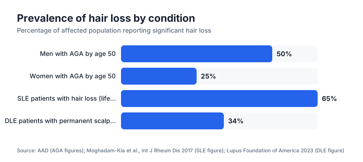
One detail worth knowing: roughly 45 to 85 percent of people with SLE report hair loss at some point during their illness, making it one of the most common outward signs of the disease [2].
What is androgenetic alopecia and what drives it?
Androgenetic alopecia (AGA) is the most common cause of hair loss on the planet, affecting roughly 50 percent of men by age 50 and about 25 percent of women by age 50 [3]. The mechanism is genetic sensitivity in follicles to dihydrotestosterone (DHT), a hormone converted from testosterone by the enzyme 5-alpha reductase. DHT gradually miniaturizes follicles over years, producing progressively finer, shorter hairs until the follicle stops making visible hair at all.
In men, loss follows the Norwood-Hamilton scale: it typically starts at the temples and crown, leaving a horseshoe of hair at the sides and back. In women, the Ludwig pattern is more common: diffuse thinning at the top of the scalp with a preserved frontal hairline.
The scalp itself looks normal. No redness. No scaling. No scarring. That is the sharpest contrast with lupus. For a closer look at the hormonal side, the DHT blocker and finasteride pages cover the pharmacology in detail.
What are the key differences between lupus hair loss and AGA?
This is where pattern recognition earns its keep. The differences are real and, once you know what to look for, fairly consistent.
| Feature | Lupus hair loss | Androgenetic alopecia |
|---|---|---|
| Pattern | Patchy (DLE) or diffuse (SLE) | Patterned: temples/crown (men), top of scalp (women) |
| Scalp appearance | Redness, scaling, scarring, follicular plugging (DLE); normal in SLE | Normal, no inflammation |
| Permanence | Permanent if scarring (DLE); reversible if no scarring (SLE) | Progressive but treatable |
| Speed of onset | Can be rapid during a flare | Slow, over years |
| Associated symptoms | Fatigue, joint pain, rash, photosensitivity | None beyond hair loss |
| Age of onset | Often 20s-40s | Starts 20s-30s in men; often post-menopause in women |
| Sex distribution | 90% of lupus patients are female | Affects both sexes; more visible in men |
The scalp is the most telling clue. If the scalp looks inflamed, scaly, or scarred, AGA is essentially off the table. AGA follicles miniaturize silently. They do not inflame the skin around them.
The "lupus hairs" seen in some SLE patients are another distinctive sign: short, broken hairs about 1 to 2 cm long along the frontal hairline, caused by hair fragility during systemic flares [2]. AGA does not produce breakage like that. It converts terminal hairs to vellus hairs through miniaturization, not fracture.
Speed matters too. AGA is measured in years. Lupus-related shedding can visibly worsen over weeks during a flare.
Which tests confirm whether hair loss is lupus or AGA?
No single symptom is definitive. A proper diagnosis usually needs two or three of the following.
Scalp biopsy. This is the gold standard for separating scarring from non-scarring loss and for identifying the inflammatory pattern specific to discoid lupus. A punch biopsy from an active lesion in DLE shows interface dermatitis, thickened basement membrane, and perifollicular inflammation with mucin deposition [4]. AGA biopsy shows miniaturized follicles with an increased vellus-to-terminal ratio and no inflammation.
Blood tests. If lupus is suspected, a rheumatologist or dermatologist will order antinuclear antibody (ANA), anti-double-stranded DNA (anti-dsDNA), complement levels (C3, C4), a complete blood count, and inflammatory markers. The American College of Rheumatology's 2019 classification criteria for SLE assign point values to these findings [5]. AGA produces no abnormal bloodwork.
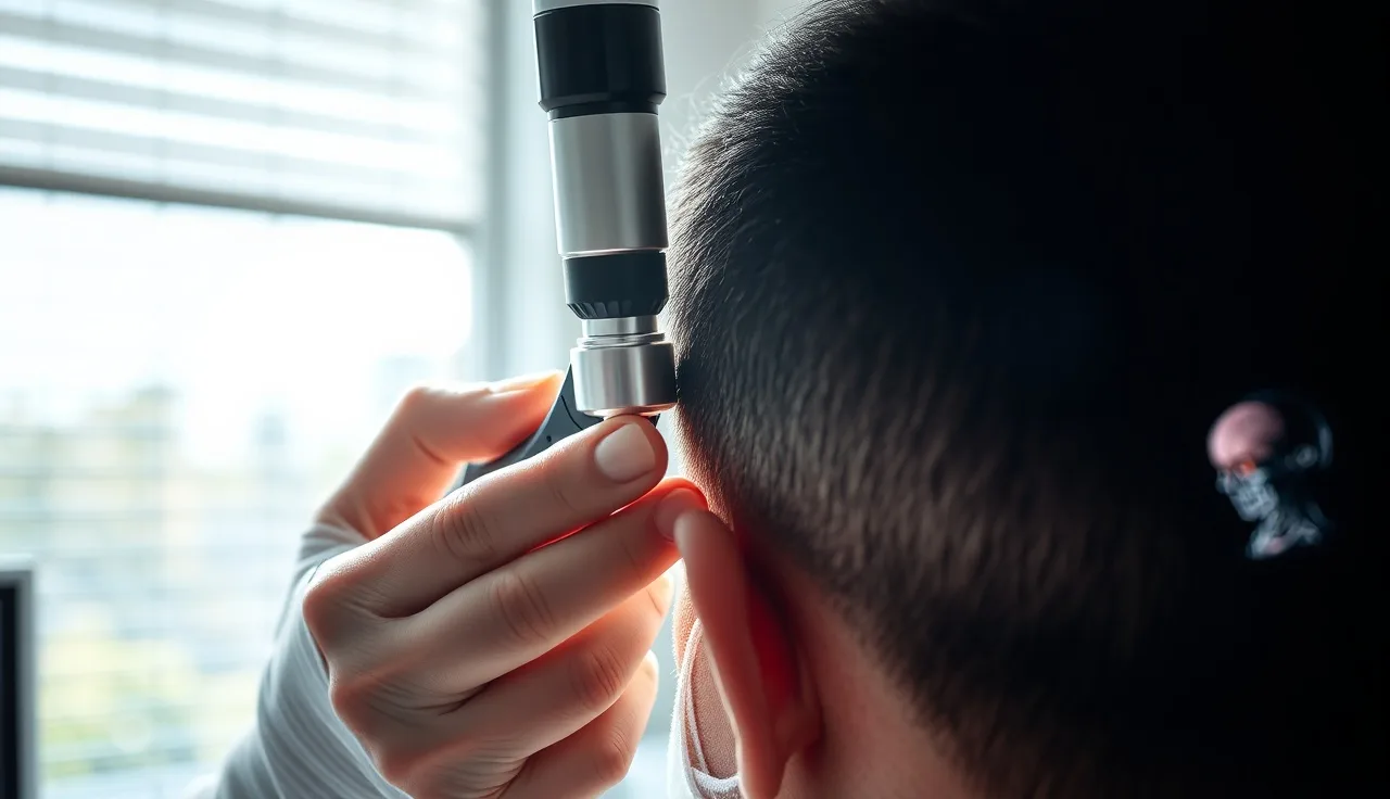
Dermoscopy (trichoscopy). A handheld or digital dermoscope lets a clinician look at the scalp surface and follicular openings without cutting. In DLE, dermoscopy shows follicular plugging, perifollicular white halos, and arborizing red vessels at scarred zones [6]. In AGA, the hallmark finding is hair diameter variability (miniaturized hairs alongside normal ones) and peripilar signs without inflammation.
Pull test. A gentle pull of 50 to 60 hairs at the scalp's periphery extracts more than 6 telogen hairs in active effluvium (including SLE-related shedding) but is typically normal in stable AGA.
If you have any systemic symptoms alongside hair loss, photosensitivity, joint swelling, unexplained fatigue, or a butterfly-shaped facial rash, skip the wait-and-see approach. A rheumatology referral alongside the dermatology visit is reasonable.
Can lupus and androgenetic alopecia occur at the same time?
Yes. They can coexist, and that combination is genuinely tricky to read. Lupus is most common in women aged 15 to 44, and women in that range can also have early AGA or the female pattern hair loss (FPHL) variant. A woman might carry the patterned thinning of FPHL as her background while developing DLE patches from lupus on top of it.
When both are present, each needs its own treatment. The lupus component is addressed through immunosuppression. The AGA component may still respond to minoxidil, which is used in both men and women. A dermatologist who does trichoscopy and also manages inflammatory scalp disease is the right specialist here.
Co-occurrence is one reason assuming AGA without a proper workup is a mistake whenever there are systemic or inflammatory signs. You could be treating the wrong condition while the other one keeps damaging follicles.
How is lupus hair loss treated?
Treatment depends entirely on which type of lupus is causing the loss.
For discoid lupus (DLE), the goal is to stop inflammation before scarring destroys more follicles. First-line treatment is typically topical or intralesional corticosteroids for localized disease [1]. If that is not enough, hydroxychloroquine (an antimalarial) is the most commonly used systemic agent. The Journal of the American Academy of Dermatology has published studies supporting hydroxychloroquine's ability to reduce DLE activity and prevent further follicle destruction [7]. Scarred areas, once set, cannot be regrown through medication. A hair transplant may be considered once the disease has stayed in remission for at least one to two years, though results vary and not every surgeon will operate on previously scarred tissue.
For SLE-related diffuse shedding (the telogen effluvium type), the primary treatment is controlling the underlying disease. As flares are managed with hydroxychloroquine, corticosteroids, or other immunosuppressants, hair usually regrows over three to six months. Topical minoxidil is sometimes added to speed regrowth, though solid trial data specific to this population is thin.
One more thing lupus patients should watch for: some drugs used to treat lupus, including certain blood pressure medications and immunosuppressants, can cause hair loss on their own. Sorting drug-induced loss from disease-induced loss takes a careful medication history.
How is androgenetic alopecia treated?
AGA is better studied than almost any other hair loss condition. Two drugs have FDA approval specifically for it.
Minoxidil. Available over the counter as a 2% or 5% topical solution or foam. The FDA approved topical minoxidil for men (5%) and women (2%) for AGA [8]. Oral minoxidil at low doses (0.25 to 1.25 mg daily for women, 2.5 to 5 mg for men) is used off-label and increasingly backed by clinical data. The minoxidil for men and oral minoxidil pages go through the evidence in detail. Minoxidil needs continuous use. Stop it and the benefit reverses within months.
Finasteride. FDA-approved at 1 mg daily for men with AGA. It blocks 5-alpha reductase, cutting scalp DHT by roughly 60 percent [9]. A five-year study published in the Journal of the American Academy of Dermatology found 48 percent of men on finasteride had visible regrowth versus 7 percent on placebo at two years. It is not FDA-approved for women and is contraindicated in women who may become pregnant.
Combination. Using both at once produces better outcomes than either alone for most men, which is why the finasteride and minoxidil combination gets recommended so often.
Hair transplants. For AGA that has outrun medical therapy, follicular unit excision (FUE) or follicular unit transplantation (FUT) can permanently restore hair in areas with enough donor supply.
None of these AGA treatments touch lupus inflammation. Using finasteride or hair loss supplements for DLE-related scarring wastes your money and your time.
What does the hairline look like in each condition?
The hairline is often the first thing people notice and worry about, so it earns its own section.
In male AGA, the receding hairline follows a predictable bilateral temple recession before creeping over the crown. The recession is smooth, symmetric, and gradual. The hair along the receding edge is usually visibly finer than hair at the back and sides.
In discoid lupus on the scalp, patches can show up anywhere, including near the hairline, but they tend to be irregular, asymmetric, and accompanied by visible scalp changes: redness, white scale, or shiny scarred skin. The patches follow no hormonal or genetic map.
In SLE-related shedding, the "lupus hairs" phenomenon produces short, broken hairs specifically at the frontal hairline. This reads differently from AGA recession because the hairline position has not actually moved. What you see is hair breakage, not follicle loss.
If you are staring at photos of your hairline trying to work out what is happening, a free scan tool like MyHairline's AI analysis can help flag the pattern and whether inflammatory signs are visible. Treat it as a starting point, not a replacement for a biopsy or blood panel.
When should you see a doctor, and which specialist?
See a doctor sooner than you think you need to. The reason is simple: in discoid lupus, every week of active inflammation risks permanent follicle loss. Waiting buys you nothing.
For straightforward patterned hair loss with no systemic symptoms and a normal-looking scalp, a board-certified dermatologist with an interest in hair loss is the right call. If you have any of the following, add a rheumatology referral or at minimum ask for lupus bloodwork: a butterfly-shaped rash across the nose and cheeks, joint pain or swelling, unusual fatigue, sensitivity to sunlight, mouth sores, or hair loss patches with visible scalp redness or scarring.
The ACR/EULAR classification criteria published in 2019 [5] give rheumatologists a systematic way to gauge lupus probability based on organ involvement and serology scores. A diagnosis of SLE requires a score of 10 or more using that system. Hair loss plus a positive ANA and one other domain finding can already push someone toward that threshold.
If cost or access is a barrier, starting with a primary care physician who can order ANA and basic labs is a reasonable first step before a specialist referral.
Are there other conditions that look like either lupus or AGA?
Yes, and this is where things get genuinely complicated. Hair loss has a long differential diagnosis, and some conditions overlap with both lupus and AGA.
Alopecia areata produces patchy loss like discoid lupus but without scalp scarring or inflammation you can see with the naked eye. Dermoscopy and biopsy separate them cleanly.
Frontal fibrosing alopecia (FFA) is a scarring alopecia that produces hairline recession resembling AGA in women, but the hairline edge shows perifollicular redness and scaling, the eyebrows and body hair are often affected too, and biopsy shows lichenoid inflammation rather than androgenetic miniaturization [10].
Telogen effluvium from any cause (illness, crash dieting, postpartum changes, iron deficiency) produces diffuse shedding that mimics SLE-related loss. The distinction depends on finding the trigger and running the right labs. The telogen effluvium page covers this in depth.
Secondary syphilis can cause a moth-eaten alopecia that looks patchy. It is less common but belongs on the differential in sexually active patients with unexplained patchy loss.
The lesson from all of this: self-diagnosing hair loss from photos or symptoms alone is unreliable. If the pattern does not fit clean male AGA in a man with no other symptoms, a proper workup is worth the time and cost.
Can hair regrow after lupus-related loss?
It depends entirely on whether scarring happened.
In active SLE without scalp scarring, hair loss is usually reversible. Once systemic disease activity is under control, most patients see meaningful regrowth within three to six months [2]. The follicles are still intact. They were pushed into a stress-response shedding phase, not destroyed.
In discoid lupus with established scarring, fibrotic tissue has replaced the follicle. No medication reverses mature scar tissue. Treating DLE early and aggressively, before it scars, is why prompt diagnosis carries so much weight.
In borderline areas where inflammation is active but scarring is incomplete, treatment can sometimes save follicles that are not yet fully gone. Another argument for moving fast.
For AGA, regrowth is possible, but keep expectations honest. Minoxidil and finasteride maintain and partially restore hair. They rarely produce dramatic regrowth in areas bald for years. The earlier treatment starts in AGA, the better the outcome, for the same reason as lupus: once a follicle is fully miniaturized and gone, medical therapy cannot bring it back. Understanding what causes hair loss in general helps set realistic expectations across all these conditions.
Sources
- American Academy of Dermatology, Lupus and hair loss page
- Moghadam-Kia S et al., 'Management of cutaneous manifestations of systemic lupus erythematosus,' International Journal of Rheumatic Diseases, 2017
- American Academy of Dermatology, Hair loss statistics
- Patel P & Werth V, 'Cutaneous lupus erythematosus: a review,' Dermatologic Clinics, 2002; PMID 12380049
- ACR/EULAR 2019 classification criteria for SLE, Arthritis & Rheumatology
- Tosti A et al., 'Dermoscopy of hair and scalp disorders,' Journal of the American Academy of Dermatology, 2008
- Kuhn A et al., 'Hydroxychloroquine in lupus: Effects on disease activity and prevention of flares,' Journal of the American Academy of Dermatology, 2020
- FDA, Rogaine (minoxidil 5%) label and approval history
- Kaufman KD et al., 'Finasteride in the treatment of men with androgenetic alopecia,' Journal of the American Academy of Dermatology, 1998; PMID 9790113
- Vañó-Galván S et al., 'Frontal fibrosing alopecia: a multicenter review of 355 patients,' Journal of the American Academy of Dermatology, 2014
- Lupus Foundation of America, About lupus prevalence statistics