
TL;DR: Assess graft survival at 12 months, not before. A good outcome shows 80 to 90% graft survival and a recipient density of 35 to 50 follicular units per cm². Track progress with monthly photos, a phototrichogram, or clinic trichoscopy. Zero growth in a zone by month 10, or heavy patchiness, means you call your surgeon.
What counts as good graft survival and density after a hair transplant?
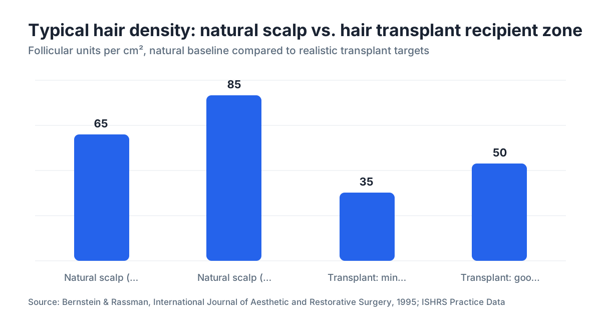
A good result means 80 to 90% of your grafts survive at 12 months, and the recipient zone holds 35 to 50 follicular units per cm². Those two numbers, survival rate and recipient density, are what every follow-up should center on. Everything else feeds into them.
The 80 to 90% survival figure is the most cited benchmark in hair restoration literature [1]. Implant 2,000 grafts and you'd expect 1,600 to 1,800 of them to keep growing hair. Survival below 70% is a poor outcome. That's a conversation with your surgeon, not a shrug.
Density is a separate measurement, and it's where expectations go sideways. The natural scalp carries roughly 65 to 85 follicular units per cm² depending on ethnicity and individual variation [2]. Transplants rarely match that. A realistic recipient target is 35 to 50 follicular units per cm², which produces the optical impression of fullness when light hits the scalp at an angle.
Whether 40 units per cm² actually looks thick depends on your hair. Caliber, color contrast with your scalp, and wave pattern all change the picture. Fine hair packs an optical punch above its number. Sparse coarse hair on a pale scalp does not.
When can you actually start evaluating results?
You cannot meaningfully judge graft quality before 10 to 12 months. The reason is biology, not marketing. Transplanted follicles shed, go quiet, then wake up on their own schedule, and the ones that lag behind can take nearly a year to show up.
The cycle runs in a predictable order: shock loss (weeks 2 to 6), dormancy (months 1 to 4), early regrowth (months 4 to 6), thickening (months 6 to 10), and near-final result (months 10 to 14) [3]. Trichoscopy studies show mean hair density keeps climbing between the 6-month and 12-month mark in most patients. That's why a photo at month 6 can look alarming while everything is fine.
Here's a practical timeline:
| Milestone | What you can evaluate |
|---|---|
| Week 1 to 2 | Crust shedding, graft "popping" (normal), wound healing |
| Month 1 to 3 | Almost nothing meaningful; shedding is expected |
| Month 4 to 6 | First fine hairs emerging; uneven density is normal here |
| Month 8 to 10 | Rough sense of coverage pattern; still thickening |
| Month 12 | First valid density assessment; good baseline for photos |
| Month 18 | Final check, especially for slow-growing coarser hair |
If a clinic calls your result final at 6 months, treat that as a red flag.
How do clinics measure graft density objectively?
Clinics use three main tools: trichoscopy, phototrichogram, and standardized macrophotography. Knowing what each one does lets you ask for the right one at your follow-up instead of accepting a vague "looks great."
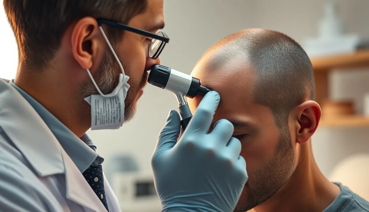
Trichoscopy (dermoscopy of the scalp) uses a polarized-lens camera magnified 20 to 70x to count hair shafts per cm². It's non-invasive, takes about 10 minutes, and gives you a number you can track [4]. A clinic that runs trichoscopy at both the pre-op consult and the 12-month visit hands you a real before-and-after.
A phototrichogram is more precise. The technician clips a small area, photographs it, waits 48 to 72 hours, then photographs again. Hairs that grew are in the anagen (active) phase. This separates growing hairs from dormant ones and produces an anagen-to-telogen ratio. It's not common outside academic centers and specialist clinics, but it's the closest thing to an objective survival count short of a biopsy.
Standardized macrophotography is the floor, the bare minimum you should expect. Your clinic shoots the same zones under the same lighting at each visit, usually with a VECTRA system or a trichoscale grid. Skip the standardization and any comparison is worthless, because angle and lighting swing apparent density hard. Ask what the photo protocol is before you book surgery.
Scalp biopsy confirms follicle presence directly but it's invasive and almost never justified for routine follow-up. It's for cases where inflammatory scarring alopecia is suspected behind graft failure.
How can you track your own progress at home?
You don't need clinical gear for basic tracking. Consistency beats precision every time.
Shoot photos on the same day each month, in the same spot, under natural daylight. Skip bathroom lighting, which shifts unpredictably and either flatters or wrecks the look. Hold the same camera-to-head distance. Take three angles: top-down, hairline straight on, and each temple at 45 degrees. Save them in dated folders and compare month to month, never day to day.
For a rough density read without tools, wet your hair and comb it straight back under bright light. The zones where scalp shows through most are your thinnest areas. Photograph those specifically. It's crude and it's reproducible, which is the point.
If you want a more structured read of your photos, the free AI scan at MyHairline can analyze scalp images for density patterns and line them up against Norwood staging. It won't replace trichoscopy. It does help you name what you're seeing before a clinic visit.
One habit pays off: note the date you first spot a new hair in a given zone. Plot those dates across your photos and you get a rough map of which graft zones fired early and which dragged. That map tells your surgeon where to look if something feels off.
What are the signs that grafts are failing or have poor quality?
Failing grafts show three signs: zero visible growth in a specific zone by month 10, heavy patchiness that ignores the natural density gradient your starting Norwood stage would predict, or a survival estimate well below 70% at the 12-month trichoscopy count. Slow growth is usually fine. These are not.
Poor hair quality in surviving grafts is a different problem. Hairs that come in very fine and stay fine, or that grow curly or kinked in ways your natural texture never did, can point to follicle trauma during harvest or implantation. This happens more with fast manual punch extraction than with slow motorized FUE or FUT strip harvesting, where the follicle is less likely to get cut.
Transection rate is the percentage of follicles damaged at harvest. Experienced FUE surgeons run around 5 to 10%; rushed or inexperienced extraction can push it past 15 to 20% [1]. That number never shows up on your invoice. Ask your surgeon for their average transection rate before you book.
Focal patchiness, where one spot stays bald while everything around it grew, can signal recipient site problems: sites angled wrong, packed too close so grafts compete for blood, or thermal damage from electrocautery misused during site creation.
Diffuse thinning of transplanted hairs that grew and then seem to miniaturize again is a separate issue. That's usually continued androgenetic alopecia hitting the native follicles mixed into the recipient area, not graft failure. This is exactly why surgeons and dermatologists push medical therapy after a hair transplant. Finasteride and minoxidil for men are the two evidence-backed options for slowing that slide.
What causes grafts to fail or grow in poorly?
Most graft failure has a traceable cause, and knowing them matters if you're weighing a second procedure or judging a clinic. Here are the common ones.
Graft handling time. Follicular units kept outside the body start losing viability, with meaningful decline after 4 to 6 hours even in chilled saline [5]. Some clinics now use ATP-enriched holding solutions such as HypoThermosol or Plasmalyte to stretch that window. Worth asking about.
Overpacking. Cram more than 40 to 50 grafts per cm² into one session and you strain the local blood supply. Grafts nearest the center of a dense cluster can die from ischemia. Good surgeons stage dense areas across multiple sessions instead of forcing it.
Technician skill. In many clinics the surgeon creates the sites while technicians handle extraction and implantation. Their skill drives transection rate and graft desiccation directly. Pre-op consults skip over this far too often.
Recipient blood supply. Patients with heavy scarring from prior surgery, or conditions that cut scalp circulation, start with less blood flow to keep grafts alive.
Post-op infection. Bacterial folliculitis in the first 4 weeks can destroy grafts. It shows up as pustules around implanted hairs and needs prompt antibiotics.
Smoking. Nicotine causes vasoconstriction that measurably drops scalp perfusion. Most surgeons ask patients to quit 2 to 4 weeks before and after surgery, and the evidence is consistent enough that it sits in ISHRS post-operative guidance [6].
How does FUE graft quality compare to FUT?
Neither technique wins on graft quality by default. The honest answer is that the surgeon matters more than the method. A well-run FUT beats a rushed FUE, and the reverse holds too.
FUT (strip surgery) removes a strip of scalp from the donor zone, and technicians dissect individual follicular units under microscopes. That dissection happens outside the body with clear visibility, which tends to keep transection low (often under 5% in skilled hands) and lets a single session harvest large graft numbers [7].
FUE extracts grafts one at a time with a circular punch, usually 0.8 to 1.0 mm. Done well, its transection rate matches FUT. Done by someone inexperienced or in a hurry, it climbs. FUE also leaves grafts out of the body longer during big sessions, which chips at viability.
From the recipient's side, graft quality per unit delivered can be similar. The donor scar differs (a linear line versus scattered small dots), recovery differs, but there's no strong evidence that 12-month density outcomes split between the techniques when equally skilled teams run them [7].
So the comparison that counts isn't FUE versus FUT. It's skilled team versus rushed team. Ask any clinic for documented graft survival data, more than a wall of before-and-after photos.
Does medical therapy after surgery affect graft outcome or density?
Yes, indirectly and significantly. Grafts taken from the permanent donor zone (usually the occipital scalp) are generally DHT-resistant. They keep the genetic programming of where they came from, so they shouldn't miniaturize from androgenetic alopecia the way native follicles do. That resistance is the whole principle behind hair transplantation.
The native hairs surrounding those grafts are not resistant. Without treatment you can land in a bad spot: transplanted hair holds while native hair keeps thinning around it, leaving an island of density in a receding sea that reads as unnatural within a few years.
Finasteride (a DHT blocker) slows that. A 5-year study by Kaufman et al. in the Journal of the American Academy of Dermatology reported that men on 1mg finasteride daily gained a mean of 107 hairs in a 1 inch² area over 5 years compared to placebo, and held higher counts throughout [8].
Minoxidil, topical or oral (see oral minoxidil), supports both native and transplanted follicles by extending the anagen phase and improving scalp blood flow. The combination of finasteride and minoxidil is the standard maintenance approach post-transplant for patients with androgenetic alopecia [11].
Skip medical therapy after a transplant and you protect the grafts while the canvas around them deteriorates. Most hair restoration surgeons will say this to your face.
What should your 12-month follow-up appointment actually include?
A real 12-month evaluation includes standardized photos, trichoscopy of both zones, a density count in your problem areas, an honest survival estimate, and a straight talk about next steps. Most clinics run one or two follow-ups at 1 and 6 months and call it closed. That's not enough.
Standardized photography of all treated zones, shot to match your pre-op conditions. This is the basic quality check and it's non-negotiable.
Trichoscopy or dermatoscopy of the recipient zone and the donor zone. Donor matters because repeated FUE can cause cumulative scarring or depletion that limits any future session.
A density estimate per cm² in your main areas of concern. If they run trichoscopy, ask for the follicular unit count per cm² in writing.
An honest read on what percentage of implanted grafts appear to have survived. Your clinic should have a record of how many grafts went into each zone.
A check on whether the result meets your pre-op expectations, and if it doesn't, what the realistic options are. That might mean a second session, a change to medical therapy, or platelet-rich plasma (PRP), which has some evidence for density improvement though a thinner evidence base than finasteride or minoxidil [9].
If your clinic doesn't offer a formal 12-month assessment, ask for one. Clinics that stand behind their work want this data as much as you do.
What if your results look patchy or thinner than expected?
Don't conclude anything before 12 months. Uneven early regrowth is common, and zones that look sparse at 7 months often fill in by month 12 as hairs thicken and more emerge from dormancy. Telogen effluvium can hit transplanted areas the same way it hits native hair, briefly adding to the apparent thinness in the early months.
Past 12 months and density genuinely looks low? Work through these steps.
Get a formal trichoscopy count. You need an objective number, not a visual impression.
Ask your surgeon to compare that count to the number of grafts implanted and estimate a survival rate. Below 70% is a legitimate outcome concern worth pressing on.
Weigh whether native hair loss has kept moving. A transplant done 18 months ago at Norwood 3 can look different now because native recession continued underneath. A medical therapy check belongs here.
Get a second opinion from a separate IAHRS or ISHRS-affiliated surgeon. An independent scalp exam carries more weight than reassurance from the clinic that did the original work.
PRP or low-level laser therapy (LLLT) are sometimes tried to support lagging grafts, though neither has strong randomized controlled trial evidence for established post-transplant cases [9].
If hair loss is still ongoing despite the transplant, it's worth reopening the receding hairline plan and any second-session decision, ideally after you've stabilized things with medical therapy first.
Can a second AI scan or online photo review give you useful information?
It gives you structure, with clear limits. An AI density analysis like the one at MyHairline can read your photos, flag low-density areas, and line your pattern up against clinical staging benchmarks. That's genuinely useful for understanding what you're looking at before or between clinic visits.
What no photo tool can tell you: actual follicular units per cm², transection damage in individual grafts, or whether a thin-looking spot has dormant follicles still to emerge. Those need physical exam or equipment.
Use photo analysis to document, track, and communicate. Print the read and bring it to your surgeon. It turns a vague "I think the crown looks thin" into a concrete starting point.
The ceiling for at-home assessment is this: you can tell whether your result sits in the normal range for your stage of recovery, and you can flag zones that stay thin so your clinic aims their evaluation there. That's a real contribution, and it's also where the tool stops.
Sources
- International Society of Hair Restoration Surgery (ISHRS), Practice Census and Outcome Data
- Bernstein RM, Rassman WR. Follicular transplantation. International Journal of Aesthetic and Restorative Surgery. 1995
- Stough DB et al. Nursing hair transplant patients. Dermatologic Surgery. 2005; via PubMed
- Rakowska A et al. Trichoscopy as a method of assessment of hair and scalp diseases. Journal of Drugs in Dermatology. 2008; via PubMed
- Cooley JE. Optimal graft care. Hair Transplant Forum International. ISHRS. 2011
- ISHRS Post-Operative Patient Care Guidelines
- Rassman WR et al. Follicular unit extraction: minimally invasive surgery for hair transplantation. Dermatologic Surgery. 2002; via PubMed
- Kaufman KD et al. Finasteride in the treatment of men with androgenetic alopecia. Journal of the American Academy of Dermatology. 1998 and 5-year update; via PubMed
- Gupta AK et al. Platelet-rich plasma as a treatment for androgenetic alopecia. Dermatologic Surgery. 2019; via PubMed
- FDA Drug Label: Finasteride 1mg (Propecia), NDA 020788
- American Academy of Dermatology (AAD), Hair Loss: Diagnosis and Treatment
- Limmer BL. Elliptical donor stereoscopically assisted micrografting as an approach to further refinement in hair transplantation. Dermatologic Surgery. 1994; via PubMed