

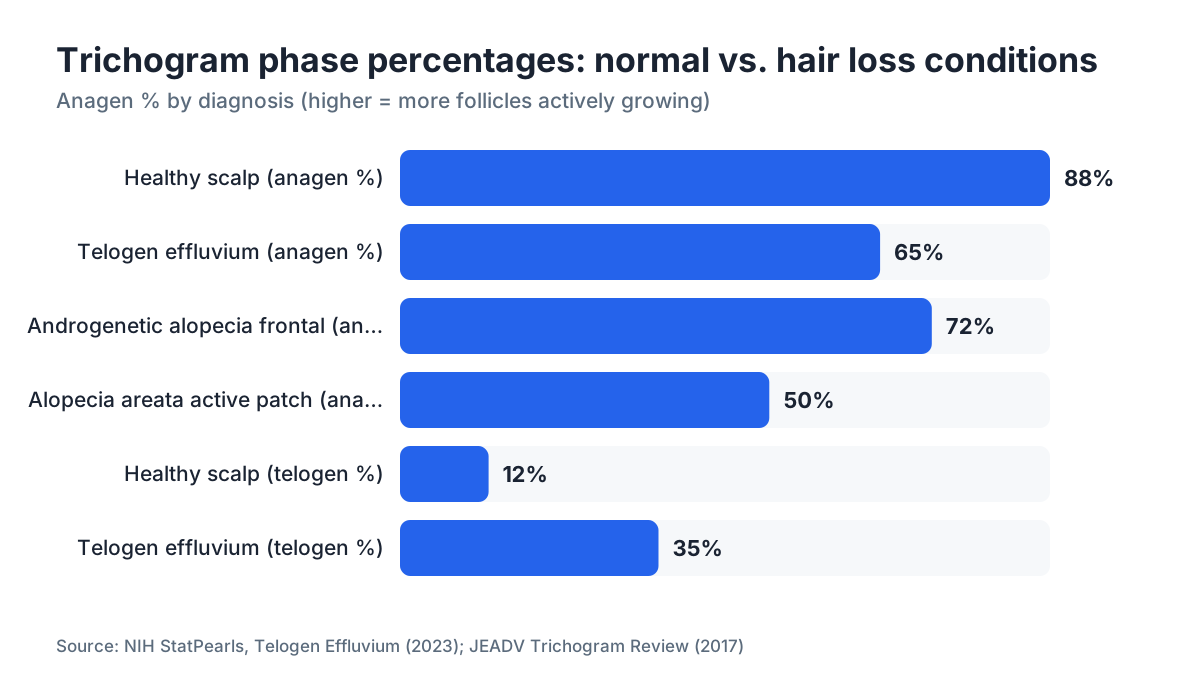
TL;DR: A trichogram pulls 50-100 hairs and sorts them under a microscope into growth (anagen), resting (telogen), and damaged (dystrophic) categories. Healthy scalps run roughly 85-90% anagen and under 15% telogen. Results above 25% telogen suggest telogen effluvium. A high dystrophic count points toward alopecia areata. Your dermatologist uses those ratios to confirm a diagnosis before prescribing treatment.
What is a trichogram and why do dermatologists order it?
A trichogram is a microscopic hair-root analysis. The clinician grips a small bundle of hairs with rubber-tipped forceps, pulls them sharply from the scalp, and mounts the roots on a glass slide. Under magnification, each hair root tells a story about where that follicle sits in its growth cycle.
Dermatologists order it when shedding looks abnormal but a visual exam alone can't pin down the cause. It's one of the few tests that gives you actual numbers: what percentage of your follicles are actively growing right now versus sitting idle. That distinction drives very different treatment paths.
The test has been used in clinical practice since the 1960s. A 2017 review in the Journal of the European Academy of Dermatology and Venereology described the trichogram as "one of the oldest and still most informative diagnostic methods for hair disorders" [1]. It's not perfect, and it depends on the operator's technique, but it's cheap, it's fast, and it gives objective data that a pull-test alone cannot.
What do the three hair phases mean in your results?
Every hair follicle cycles through three phases, and your trichogram counts which phase each plucked hair was in when it was pulled.
Anagen (growth phase): The follicle is actively making a hair shaft. Anagen roots look long, pigmented, and sheathed in a white inner root sheath when viewed under the microscope. Normal scalp hair spends 2-7 years in anagen, which is why most hairs on a healthy head are in this phase.
Telogen (resting/shedding phase): The follicle has stopped producing fiber and is preparing to shed. Telogen roots look club-shaped, dry, and unpigmented. They detach easily. A hair in telogen will shed on its own within weeks whether you pluck it or not.
Catagen (transition phase): The brief transition between anagen and telogen, lasting only 2-3 weeks. Catagen hairs are rarely captured in a standard trichogram because so few follicles are in this phase at any moment. Most labs don't report catagen separately. They fold it into the anagen count or flag it as a separate note.
Dystrophic hairs are a fourth category some labs report. These are broken anagen hairs with abnormal, tapered, or fractured roots. They signal that the follicle was forced out of active growth prematurely, which matters a great deal for diagnosis.
Think of the anagen-to-telogen ratio as the core signal. Everything else is context.
What are the normal reference ranges for a trichogram?
The accepted normal values in the literature are [1][2]:
| Metric | Normal range | Concern threshold |
|---|---|---|
| Anagen % | 85 - 90% | Below 80% |
| Telogen % | 10 - 15% | Above 20-25% |
| Dystrophic hairs | < 2% | Above 5% |
| Catagen % | < 3% | Rarely flagged alone |
These numbers come from studies using standardized technique: hairs plucked from a defined scalp area, usually the mid-frontal and occipital regions, after patients have not washed their hair for 24-48 hours (washing removes loose telogen hairs and skews the count).
A few cautions about these ranges. They are population averages, and some labs use slightly different cutoffs. Seasonal variation is real: a 2009 study in the British Journal of Dermatology found telogen rates naturally peak in autumn [3]. If your result says 18% telogen in October, that deserves a different conversation than 18% telogen in March. Always ask which scalp region was sampled, because the frontal scalp in androgenetic alopecia can run higher telogen counts than the back of the head, which is why dermatologists often sample both and compare.
How do trichogram results differ by condition?
The numbers shift in characteristic patterns depending on what's going wrong.
Telogen effluvium: Telogen percentage climbs above 25%, often reaching 30-50% in acute cases. Anagen roots are normal-looking. Dystrophic count stays low. This pattern means follicles were pushed into resting phase by a systemic trigger (severe illness, crash diet, major surgery, postpartum hormonal shift). You can read a full breakdown of triggers at telogen effluvium.
Androgenetic alopecia (pattern hair loss): The trichogram on the frontal or vertex scalp shows a lower anagen percentage than the occipital scalp. The occipital region acts as an internal control. You might see 70% anagen frontally versus 87% occipitally. The frontal hairs also tend to have thinner shaft diameters, which a trichoscopy (dermoscopy of the scalp) quantifies better than a basic trichogram. This regional gap is the fingerprint of androgenetic loss. Understanding what causes hair loss in pattern baldness, primarily DHT, gives context for why the front goes first.
Alopecia areata: Dystrophic (broken anagen) hairs shoot up, sometimes above 30%. These are "exclamation mark" hairs broken off at the root by the immune attack on the follicle. The total anagen percentage may also be very low in active patches. This is a clinically distinct picture from telogen effluvium.
Normal or no diagnosis: If your numbers land in the normal ranges but you still feel you're losing hair, the trichogram is not the final word. Hair density, scalp coverage, and miniaturization patterns matter too. The test samples roughly 50-100 hairs from a small area. It doesn't capture the full picture of your scalp.
Here's the practical read: anagen low plus high dystrophic means something is actively damaging follicles now. Anagen normal but telogen elevated means follicles were healthy but got pushed to rest weeks or months ago. Both patterns need treatment. They just need different treatment.
How is the test performed, and does the technique change your results?
Technique matters more than most patients realize. The standard protocol, described in the textbook Trichology by Sinclair and colleagues, calls for the following:
- No shampooing for 24-48 hours before the test. Washing rinses out loose telogen hairs, artificially inflating the anagen percentage.
- Clamp 20-30 hairs at once with rubber-tipped forceps. Pull fast, in the direction of hair growth.
- Mount roots on a glass slide in a mounting medium and examine at 40-100x magnification.
- Count at least 50 roots per site for statistical reliability. Many labs count 100.
Where on the scalp hairs are pulled matters. Frontal, vertex, and occipital sites each give different baselines. If the lab report doesn't state the sampling location, ask. A single frontal result without an occipital comparison is harder to interpret.
Some clinics now use video trichoscopy, which measures hair shaft diameter and follicular unit density alongside phase counts. That adds useful information but is a different test. A phototrichogram (trichogram plus sequential photography) can track the same follicles over days to confirm which phase they're in, and it's more accurate than a single-point trichogram for borderline cases [2].
Home trichogram kits exist and are generally not reliable. Root preservation is time-sensitive, magnification matters, and phase classification requires training. If you want a real result, get it done in a dermatology clinic.
What does a high telogen percentage actually mean for treatment?
A telogen percentage above 25% is the trichogram's most common abnormal finding, and it means your follicles have already been through a stressful event. The shedding you're seeing now started 8-12 weeks before your worst symptoms, because that's how long it takes follicles to cycle from early telogen into actual shedding.
For most people with telogen effluvium, the first intervention is identifying and removing the trigger: correct iron-deficiency anemia, stop a culprit medication, address thyroid disease, or wait out postpartum recovery. Follicles in telogen are not dead. They're dormant. Most will return to anagen on their own within 3-6 months of trigger removal.
If the telogen percentage is persistently elevated over multiple tests or if it's combined with the frontal-versus-occipital gap of androgenetic alopecia, that changes the picture. In that case, a DHT blocker like finasteride or a topical agent like minoxidil for men may be appropriate. The trichogram result, combined with clinical exam, guides which route to take.
One thing worth saying plainly: a trichogram tells you about the current state of follicles. It doesn't predict whether a given treatment will work for you. That takes 6-12 months of treatment and follow-up evaluation.
If you want a starting point before your dermatologist appointment, the free AI scan at MyHairline can analyze your scalp photos and flag pattern characteristics worth discussing with your doctor.
What does a high dystrophic hair count mean?
Dystrophic hairs are anagen hairs that broke before they could fully exit. Under the microscope, they look like a frayed rope end rather than a clean root. The follicle was clearly in growth phase, but something cut the process short.
A dystrophic rate above 5% raises suspicion for alopecia areata, the autoimmune condition where the body's T-cells attack hair follicles. In active alopecia areata, the rate can exceed 25-30%, with the classic "exclamation mark" morphology (tapered, narrow at the base where it broke off near the scalp). The American Academy of Dermatology identifies alopecia areata as affecting roughly 2% of people at some point in their lives [4].
High dystrophic counts can also appear in trichotillomania (compulsive hair pulling) and in anagen effluvium from chemotherapy, where the drug arrests rapidly dividing cells in the hair matrix and causes mass anagen shedding.
Telling these apart requires clinical context. A dermatologist will look at the pattern of hair loss on the scalp, the morphology under trichoscopy, and possibly a scalp biopsy before landing on a diagnosis. A trichogram is a clue, not a conclusion.
How does a trichogram compare to other hair loss tests?
Several tests are available, and they answer different questions.
| Test | What it measures | Best for | Limitation |
|---|---|---|---|
| Trichogram | Anagen/telogen/dystrophic ratio | Confirming telogen effluvium, alopecia areata | Operator-dependent; samples small area |
| Trichoscopy / dermoscopy | Hair shaft diameter, follicular unit density, scalp skin signs | Androgenetic alopecia pattern; scalp disease | Doesn't give phase ratios |
| Scalp biopsy | Follicle histology, immune infiltrate, fibrosis | Scarring alopecias; ambiguous trichoscopy | Invasive; leaves small scar |
| Pull test | Very rough estimate of shedding rate | Quick bedside screen | High false positive/negative rate |
| Blood panel (ferritin, TSH, CBC) | Systemic causes of shedding | Ruling out iron, thyroid, or CBC abnormalities | Doesn't evaluate follicles directly |
The trichogram sits between the quick pull-test and the more invasive biopsy. For suspected telogen effluvium or alopecia areata, it's often all you need. For suspected scarring alopecia (lichen planopilaris, frontal fibrosing alopecia), a biopsy is usually required because the trichogram won't show fibrosis or immune infiltrates around the follicle.
Most dermatologists combine at least two tests. A trichogram plus trichoscopy, or a trichogram plus a blood panel, gives a much more complete picture than either alone.
How do you prepare for a trichogram to get accurate results?
Preparation directly affects accuracy. Here's what changes numbers in a meaningful way:
Don't wash your hair 24-48 hours before. Shampooing dislodges loose telogen hairs. If you shower right before the test, the lab loses the telogen hairs that would have been captured, and your anagen percentage looks falsely high.
Avoid vigorous scalp massage or brushing the day before. Same reason: mechanical loosening removes resting hairs before the forceps can capture them.
Don't apply heavy styling products. They can obscure the root morphology under the microscope.
Tell your doctor about medications. Minoxidil can shift follicles toward anagen, potentially masking a high-telogen picture. Some clinicians ask patients to pause topical minoxidil for a defined period before testing, though there is no universal consensus on how long.
Bring records of recent stressors. Major illness, surgery, weight loss, pregnancy, or a crash diet in the past 3-6 months can explain a high telogen count without any ongoing scalp pathology. The trichogram can't flag the cause, only the effect. You need to provide that context.
If you're tracking treatment progress over time, try to standardize conditions between tests: same preparation, same time of year (to reduce seasonal variation), same technician if possible.
Can you interpret a trichogram report at home, or do you need a dermatologist?
You can understand the numbers. Interpreting what they mean for your specific situation is harder without clinical training.
The report will typically list anagen %, telogen %, and sometimes dystrophic % for one or more scalp regions. Comparing those numbers to the reference ranges in this article gives you a reasonable sense of whether your result is abnormal. If your telogen is 32%, you know that's well above the 10-15% normal range. That's useful information.
What you can't do at home is integrate those numbers with the pattern of your hair loss, the visual appearance of your scalp under magnification, the texture and miniaturization of remaining hairs, or your blood work. A 32% telogen reading in a 28-year-old woman six months postpartum is a completely different clinical situation than a 32% telogen reading in a 45-year-old man with a receding hairline.
If your report came from a dermatologist visit and you're trying to understand it before your follow-up, this article gives you the vocabulary. If you had the test done through a hair clinic that didn't provide much explanation, it's worth asking specifically: what site was sampled, how many hairs were counted, and how does the result compare to age-matched norms. Those questions will get you more useful answers than the raw percentages alone.
For an initial, no-cost read of your scalp photos, MyHairline's free AI scan can identify visible pattern characteristics before you even book a clinical test.
What happens after you get your trichogram results?
The trichogram is a starting point for treatment planning, not the endpoint.
If results point to telogen effluvium, your dermatologist will likely order blood work to find the trigger: ferritin (iron stores), TSH (thyroid), complete blood count, and sometimes zinc or vitamin D. Treating the underlying deficiency is the primary move. Hair regrowth after trigger removal typically takes 3-6 months.
If results suggest androgenetic alopecia confirmed by regional comparison, you're looking at a longer conversation about finasteride and minoxidil together, or one versus the other, depending on how much loss has already occurred. It may also be worth checking minoxidil side effects before committing to long-term topical or oral minoxidil. For people with significant established loss, hair transplant evaluation may eventually enter the picture.
If results suggest alopecia areata with high dystrophic counts, the treatment discussion shifts to immunomodulatory options: topical or intralesional corticosteroids, topical immunotherapy, or the newer JAK inhibitors.
A follow-up trichogram 6-12 months into treatment can objectively measure whether anagen percentage has improved. That's one of the test's real advantages over photography alone: numbers don't lie about trend direction.
Sources
- Springer: Trichoscopy: A New Method for Diagnosing Hair and Scalp Diseases (Rudnicka et al., 2012)
- American Academy of Dermatology: Alopecia Areata Overview
- National Institutes of Health (NIH) MedlinePlus: Hair loss overview
- FDA: Minoxidil drug label (DailyMed, NLM)
- PubMed / Journal of Investigative Dermatology: Stenn & Paus, Controls of Hair Follicle Cycling 2001
- PubMed / Dermatology Practical & Conceptual: Trichogram interpretation and standardization
- National Library of Medicine (NLM) / StatPearls: Telogen Effluvium
- PubMed / Clinical, Cosmetic and Investigational Dermatology: Trüeb, Systematic approach to hair loss in women 2010
- American Academy of Dermatology: Alopecia Areata Treatment