
TL;DR: Telogen effluvium (TE) is diffuse shedding triggered by a physiological stressor. Blood tests don't diagnose TE directly, but they identify the underlying cause in most cases. The core panel covers ferritin, TSH, complete blood count, and vitamin D. Depending on your history, zinc, B12, folate, sex hormones, and ANA may be added. Treating the deficiency usually stops shedding within 3 to 6 months.
What are blood tests actually trying to find in telogen effluvium?
Telogen effluvium is not a disease with a blood marker. There is no "TE level" a lab can measure. What blood tests do is hunt for the trigger underneath it, the metabolic, hormonal, or nutritional disruption that pushed hair follicles out of the growth phase en masse.
That distinction matters. Doctors sometimes hand patients a normal lab report and say "nothing is wrong." But a normal CBC doesn't mean TE isn't happening; it means anemia probably isn't the cause. The job of the workup is to walk through a checklist of known, reversible triggers until you find the one (or two, because multiple deficiencies can stack) that fits.
The American Academy of Dermatology lists nutritional deficiencies, thyroid disease, iron deficiency, and hormonal shifts among the leading correctable causes of diffuse hair shedding. [1] Each one shows up in blood, which is why a targeted panel is the first clinical step after a good history and scalp exam.
If you want the broader picture of what sets TE in motion before you head to a lab, telogen effluvium breaks down triggers, timelines, and what normal shedding versus pathological shedding actually looks like.
Which blood tests should be ordered for telogen effluvium?
The panel most dermatologists start with covers four categories: iron stores, thyroid function, a general hematological picture, and vitamin D. Beyond that, your personal history drives what gets added.
Core panel
| Test | What it measures | Why it matters for TE |
|---|---|---|
| Ferritin | Stored iron | The most sensitive iron marker for hair loss; serum iron misses early depletion |
| TSH | Thyroid-stimulating hormone | Both hypo- and hyperthyroidism cause diffuse shedding |
| Complete blood count (CBC) | Red cells, white cells, platelets | Flags anemia, infection, or inflammatory signals |
| 25-OH vitamin D | Circulating vitamin D | Deficiency associated with hair cycling disruption [2] |
Commonly added tests
| Test | Add if... |
|---|---|
| Free T3, Free T4 | TSH is borderline or symptoms don't match a normal TSH |
| Serum zinc | Diet is restricted, or you have GI malabsorption |
| Serum B12 and folate | Vegan/vegetarian diet, fatigue, or macrocytic cells on CBC |
| Total testosterone, free testosterone, DHEA-S | Female patient with signs of androgen excess (acne, irregular cycles) |
| Prolactin | Female patient with irregular cycles or galactorrhea |
| ANA (antinuclear antibody) | Suspicion of lupus or autoimmune alopecia |
| CRP or ESR | Chronic inflammatory state suspected |
| HbA1c or fasting glucose | Metabolic syndrome or PCOS on the differential |
| Complete metabolic panel (CMP) | Protein malnutrition, liver or kidney disease |
A review in the Journal of the American Academy of Dermatology noted that ferritin, TSH, and CBC together identify a correctable cause in a large share of TE patients, though the authors acknowledged that exact prevalence data vary across study populations. [3] That's the honest version: no single study has perfectly characterized how often each trigger appears, but the clinical consensus is steady.
What ferritin level is considered low for hair loss?
This is where patients and doctors genuinely disagree, and the debate is worth understanding.
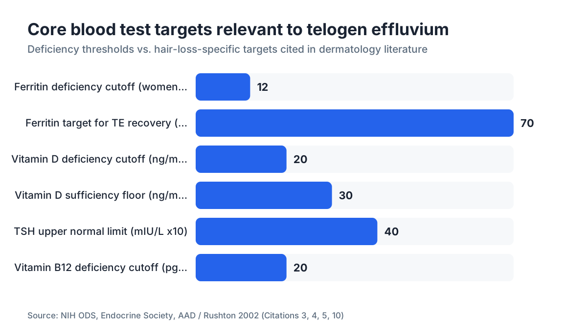
Standard lab reference ranges flag ferritin as "low" below 12 to 15 ng/mL for women and below about 20 ng/mL for men. Those cutoffs were set to prevent anemia, not to protect hair. Several dermatology researchers have argued that hair follicles need higher ferritin to run normally. A widely cited threshold in the hair loss literature is 30 ng/mL, with some clinicians repleting to 70 ng/mL or higher before calling iron stores adequate for recovery. [3]
Here's the honest position. The exact ferritin target that reliably stops TE is not pinned down by a randomized trial. What is clear: a ferritin of 10 ng/mL is almost certainly contributing to shedding, a ferritin of 80 ng/mL almost certainly is not, and the 30 to 70 ng/mL band is where clinical judgment lives.
Women of reproductive age carry the highest risk of low ferritin because of menstrual blood loss. [10] If you're in that group and your ferritin comes back at, say, 18 ng/mL with a lab report that says "normal," it's fair to ask your doctor whether iron supplementation is worth trying anyway. The downside is modest (GI upset is the main one). The upside is a reversible cause of your hair loss.
Don't supplement iron without testing first. Iron overload is harmful, and hemochromatosis patients exist. [10]
How does thyroid function cause telogen effluvium and what does the test show?
Both an underactive thyroid (hypothyroidism) and an overactive one (hyperthyroidism) disrupt the hair cycle. The mechanism differs. Hypothyroidism slows cellular metabolism across the board, shortening the anagen (growth) phase. Hyperthyroidism speeds up cycling in a way that can push large cohorts of follicles into telogen at once.
TSH is the correct first test. The pituitary secretes TSH in inverse proportion to circulating thyroid hormone, so a high TSH means the thyroid is underperforming and the pituitary is pushing it to work harder. A low TSH means the thyroid is running hot. Normal range is roughly 0.4 to 4.0 mIU/L by most U.S. laboratory standards, though clinicians still argue over whether the upper limit should be 2.5 or 3.0 for symptomatic patients. [4]
If TSH is abnormal, free T3 and free T4 clarify whether the problem is primary thyroid disease or a pituitary issue. Thyroid antibodies (anti-TPO, anti-thyroglobulin) identify Hashimoto's thyroiditis, an autoimmune condition that often causes subclinical hypothyroidism with shedding even when TSH sits in the "normal" range.
The good news. Once thyroid disease is treated and levels normalize, TE typically resolves. Expect a lag of 3 to 6 months after normalization before you see regrowth, because the follicle cycle moves slowly.
Does vitamin D deficiency cause hair loss and will a blood test catch it?
Yes on both counts. Vitamin D receptors sit inside the hair follicle, and research suggests vitamin D signaling matters for moving follicles through the anagen phase properly. [2]
Serum 25-hydroxyvitamin D (25-OH D) is the correct test. The Endocrine Society defines deficiency as below 20 ng/mL (50 nmol/L) and insufficiency as 20 to 29 ng/mL. [5] In practice, many hair loss specialists aim for at least 40 ng/mL in TE patients, though that specific threshold isn't yet backed by a clean interventional trial.
Vitamin D deficiency is common, especially in northern latitudes, in people with darker skin tones, and in people who spend most of their day indoors. A 2013 study in Skin Pharmacology and Physiology found significantly lower vitamin D levels in women with chronic telogen effluvium compared to controls, which supports the association even if causation isn't nailed down. [6]
Replacing vitamin D is cheap and simple. The question is how much. Standard maintenance runs 1,500 to 2,000 IU daily, but repleting frank deficiency may take 50,000 IU weekly (prescription D2 or high-dose D3) for 8 to 12 weeks. Retest at 3 months.
What role do sex hormones play and when should they be tested?
For women with TE, hormonal triggers are common and underdiagnosed. Postpartum hair loss is the textbook case: estrogen drops sharply after delivery, shifting a large cohort of follicles out of the growth phase. That's normal physiology and usually self-resolving. But other hormonal states can cause TE that won't clear without help: stopping hormonal birth control, perimenopausal estrogen swings, polycystic ovary syndrome (PCOS), and hyperprolactinemia.
Tests to consider in women: total and free testosterone, DHEA-S (an adrenal androgen marker), LH, FSH, estradiol, and prolactin. If PCOS is on the differential, adding fasting insulin and glucose makes sense. what causes hair loss covers the androgen pathway in more detail, including how elevated DHT drives a pattern that can look like TE but isn't.
For men, sex hormone testing is rarely the first move for TE, but low testosterone and elevated cortisol (from chronic physical or psychological stress) are legitimate contributors. A morning cortisol or DHEA-S is worth considering when the history points to adrenal stress.
Keep one thing straight. Androgenetic alopecia (pattern hair loss) and TE can coexist. A woman can have low ferritin triggering a TE shed on top of underlying female pattern hair loss. The blood work addresses the TE component. The androgen-pattern component may need separate treatment.
Can protein deficiency or B vitamin deficiencies show up on blood tests for TE?
Hair is almost entirely keratin protein. Severe protein restriction or malabsorption can absolutely cause shedding, though you see it more in eating disorders, post-bariatric surgery patients, or people on extreme elimination diets than in the general population.
A complete metabolic panel (CMP) includes albumin and total protein. These are blunt instruments. Albumin falls only in significant protein-energy malnutrition, so a normal albumin doesn't rule out the protein inadequacy that might still be starving your follicles. Prealbumin is more sensitive but rarely ordered routinely.
For B vitamins, the ones that matter are B12, folate (B9), and biotin (B7). Serum B12 below 200 pg/mL is generally considered deficient; the grey zone of 200 to 300 pg/mL is clinically debated. [11] Folate deficiency shows up on CBC as macrocytic red cells before it shows as a low folate level.
Biotin deserves a warning. It's heavily marketed for hair growth, and biotin deficiency genuinely does cause hair loss. But frank biotin deficiency is rare in anyone eating a normal diet. The FDA has issued guidance that high-dose biotin supplements can interfere with many laboratory assays, including thyroid tests and troponin, producing falsely normal or falsely abnormal results. [7] If you take biotin, stop it at least 48 to 72 hours before your blood draw and tell the lab. This one gets overlooked constantly.
For more on which supplements earn their place and which don't, hair loss supplements covers the evidence quality without the hype.
Should you test for autoimmune conditions when investigating TE?
Sometimes yes. Diffuse hair shedding is a known feature of systemic lupus erythematosus (SLE). If you have other signs, joint pain, skin rashes (particularly the butterfly rash across the cheeks), fatigue, and oral ulcers, an antinuclear antibody (ANA) test is warranted. A positive ANA with a titer above 1:80 in that clinical context sends you toward rheumatology. [8]
Autoimmune thyroiditis (Hashimoto's) sits in its own lane. As noted above, anti-TPO antibodies can explain thyroid-related TE even when TSH is technically normal. This overlap gets missed more than it should.
Alopecia areata is a different animal. It's autoimmune, but it causes patchy, not diffuse, hair loss, and it shows no specific blood marker. It gets confused with TE. If there are smooth, round bare patches rather than general thinning, the diagnosis is different and the blood workup is less useful.
How do you read the results and decide what to treat?
Getting the results is step one. The harder step is reading them against your hair loss, more than against the population reference range.
A few practical points.
Reference ranges are statistical, not biological law. They cover the middle 95% of a tested population, not the optimal level for every biological process. Ferritin of 14 ng/mL is "normal" by many labs but almost certainly inadequate for follicle function in a woman shedding 200 hairs a day.
Mild deficiencies stack. A ferritin of 22 ng/mL, a vitamin D of 18 ng/mL, and a TSH creeping toward 3.8 might together cross the threshold for triggering TE, even though each finding looks borderline on its own. Treating all three at once is reasonable.
Timing matters. Blood tests reflect current status. If you crashed your ferritin six months ago (after a heavy illness or surgery), had TE peak at four months, and are now recovering on iron, your ferritin might already be back in range by the time you test. The history of what happened three to six months before shedding started matters as much as the labs.
Once you've found a cause and treated it, expect a lag before you see anything. Hair grows about half an inch a month, and the follicle has to finish its cycle. Most people see meaningful regrowth three to six months after the trigger is removed. telogen effluvium has more on what the recovery timeline looks like.
What happens if all the blood tests come back normal?
This is frustrating and more common than it should be. Normal labs do not mean nothing is wrong. They mean the panel you ran didn't find the cause.
A few possibilities.
The trigger was in the past. TE runs a 3-month lag from trigger to peak shedding. [12] If you had major surgery in January and your labs are drawn in May after shedding started, your ferritin may have recovered by then. The trigger was real. You just can't see it in the blood anymore.
The test chosen was too blunt. Serum iron instead of ferritin misses early iron depletion. Total T4 instead of free T4 misses protein-binding interference. If your workup was limited, ask for the more sensitive markers.
The cause is physical or emotional stress, which doesn't show in routine labs. Major surgery, a severe illness, a traumatic life event, rapid weight loss, all known TE triggers that leave no blood signature six months later.
The diagnosis might need a second look. A scalp biopsy can confirm TE histologically when the clinical picture is unclear. It can also separate TE from early androgenetic alopecia, which looks similar on inspection but has different treatment implications. what causes hair loss covers how dermatologists tell the two apart.
If you want a quick way to document your current shedding pattern before seeing a specialist, the free AI analysis at MyHairline can compare your hairline and density over time, giving you visual evidence to bring to the appointment instead of relying on memory.
What should you do with the results before starting any treatment?
First, bring them to a dermatologist or a physician comfortable with hair loss, more than a GP who ran the panel as a favor. Hair loss workups need pattern recognition across several mild abnormalities, and a normal CBC alone does not close the case.
Second, fix identified deficiencies before jumping to hair-specific treatments like minoxidil. Minoxidil can help hold onto hair while a deficiency is corrected, but it doesn't fix the deficiency. Using it without addressing the root cause is treating a symptom. minoxidil for men explains how minoxidil works so you can decide whether adding it makes sense for you.
Third, if the TE is resolving but you're worried about an androgenetic pattern that may have been unmasked, that's a separate conversation about finasteride or finasteride and minoxidil combination therapy. Those are long-term medications with their own decision framework.
Finally, retest. After treating a deficiency for 3 to 4 months, check the relevant markers again to confirm correction, more than symptom improvement. A ferritin that climbed from 10 to 30 ng/mL is progress; if the target is 70 ng/mL, you may need to keep supplementing.
For most people with TE from a single correctable trigger, the prognosis is genuinely good. Hair that shed from iron deficiency, thyroid disease, or vitamin D deficiency usually grows back fully once levels normalize. That's one of the more reliable statements in all of hair medicine, and it should be reassuring.
How much do these blood tests cost and does insurance cover them?
Costs swing hard by lab, location, and insurance status. The following are approximate cash-pay prices at common U.S. commercial labs as of 2024 to 2025, before insurance adjustment.
| Test | Approximate cash price (USD) |
|---|---|
| Ferritin | $20 to $45 |
| TSH | $20 to $40 |
| Complete blood count | $15 to $35 |
| 25-OH vitamin D | $40 to $80 |
| Complete metabolic panel | $25 to $50 |
| Free T3, Free T4 | $30 to $60 each |
| Total testosterone | $30 to $60 |
| Serum zinc | $25 to $45 |
| Serum B12 + folate | $30 to $60 |
| ANA | $30 to $60 |
A core TE panel (ferritin, TSH, CBC, vitamin D) usually runs $95 to $200 out-of-pocket at a direct-access lab like LabCorp or Quest Diagnostics. With insurance, these tests are typically covered when a physician orders them for a documented medical reason (hair loss counts). Pre-authorization is rarely required for basic metabolic panels.
Ordering through a dermatologist rather than requesting the tests yourself often makes coverage smoother, since the clinical indication is documented in a specialist's chart note. If cost is a barrier, direct-to-consumer lab services let you order and pay for panels online; the prices above reflect that market. The federal No Surprises Act and state surprise billing laws may apply to lab services billed through hospital systems. [9]
Sources
- American Academy of Dermatology, Hair Loss Overview
- Molecular Endocrinology, Amor KT et al. (2010) - Vitamin D and the hair follicle
- Journal of the American Academy of Dermatology, Rushton DH (2002) - Nutritional factors and hair loss
- American Thyroid Association, Hypothyroidism Brochure
- Endocrine Society, Vitamin D Deficiency Clinical Practice Guideline
- Skin Pharmacology and Physiology, Rasheed H et al. (2013) - Serum ferritin and vitamin D in female hair loss
- U.S. Food and Drug Administration, Biotin Interference with Lab Tests Safety Communication
- American College of Rheumatology, Lupus (SLE) Resource Center
- Centers for Medicare and Medicaid Services, No Surprises Act Overview
- National Institutes of Health Office of Dietary Supplements, Iron Fact Sheet
- National Institutes of Health Office of Dietary Supplements, Vitamin B12 Fact Sheet
- Journal of the American Academy of Dermatology, Headington JT (1993) - Telogen effluvium: new concepts