
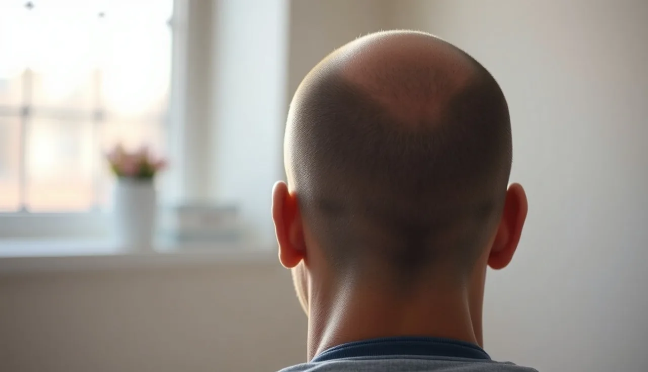
TL;DR: After FUE, each extracted follicle leaves a tiny round scar roughly 0.8 to 1.0 mm wide. At 12 months most patients show no visible scarring at normal viewing distance, provided a skilled surgeon kept extraction density below 20 to 25% of the donor zone. Thinning, wide scars, or visible pitting are signs of over-harvesting or poor technique, not normal outcomes.
What actually happens to the donor scalp when follicles are removed?
A circular punch, usually 0.8 to 1.0 mm across, rotates into the scalp around each follicular unit, and the graft gets pulled free [1]. What's left is a small open wound running the full thickness of the dermis. That wound closes by secondary intention, meaning no suture goes in. The body just seals it on its own.
Surface closure takes about 7 to 10 days. The dermis keeps remodeling for several months after that [2]. During remodeling, collagen fills in to replace the extracted tissue. The end result is a small round scar, either depigmented or skin-toned, and in most cases smaller than the original punch because the surrounding tissue contracts slightly as it heals.
Each graft site heals on its own, independent of the ones beside it. That matters. The final look depends almost entirely on two things: how close together those punch sites are, and how cleanly each wound closes. Grafts punched too close can cause two wounds to merge, which leaves an oblong or irregular scar instead of a neat round one. That merging is one of the clearest signs of poor technique.
What does the donor area look like in the first few weeks after FUE?
The first 24 to 48 hours are the roughest cosmetically. The back and sides show hundreds of tiny red dots, each ringed by a small crust. Some patients get mild swelling across the occipital area, and there can be dried blood if the hair was shaved short. All of it is expected.
By day 5 to 7, the crusts start lifting on their own. Most clinics tell patients to gently wet the scalp in the shower around day 3 and to pat, not rub, the area dry. Scratching or picking crusts off early can widen individual scars. That is one of the few post-op behaviors that actually changes the long-term result.
By the end of week two, the surface looks mostly healed to a casual observer. The dots fade from red to pink. If hair was shaved for the procedure, regrowing stubble does a lot of cosmetic work at this stage. Even 2 to 3 mm of growth starts to conceal the sites.
How visible are FUE donor scars at 3, 6, and 12 months?
At 3 months the scars are still remodeling. They can look slightly more visible than they eventually will because the collagen hasn't matured yet. Some patients notice the sites look a little shiny or paler than the surrounding scalp. Normal, and temporary.
At 6 months most of the discoloration has resolved. The scars are now white or skin-toned, and at a viewing distance past 30 cm they blend into the scalp when hair is any length longer than a grade-1 clipper cut (roughly 3 mm).
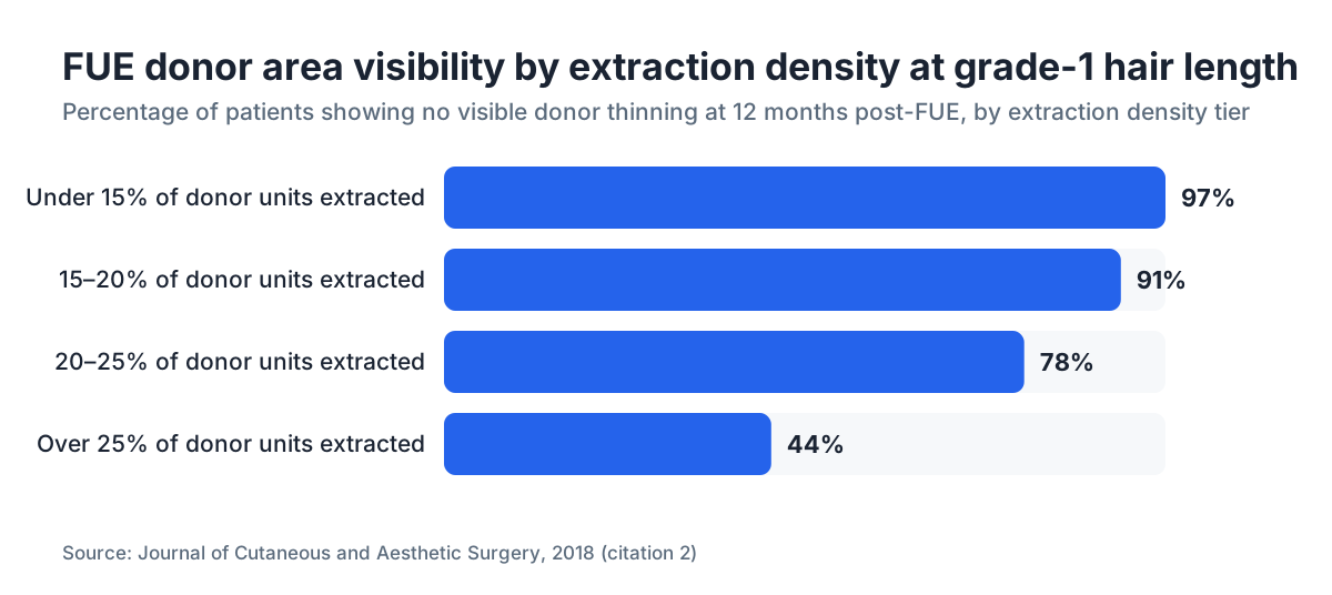
By 12 months the remodeling is largely done. A study in the Journal of Cutaneous and Aesthetic Surgery evaluated donor sites at one year and found that small punch sizes (under 1 mm) combined with extraction densities under 25% produced cosmetically acceptable results in the large majority of cases, with noticeable scarring limited to patients where over-harvesting had happened [2]. The scars don't vanish. Each one is a permanent tiny mark. But at normal hair lengths they aren't detectable without magnification.
The honest caveat: healing varies person to person. Patients who form hypertrophic scars elsewhere on their body carry a slightly higher risk of the same in the donor area, though the scalp tends to scar more quietly than the chest or back.
What is "normal" donor density loss after FUE, and what counts as over-harvesting?
The adult male donor area holds roughly 6,000 to 8,000 follicular units, and that number swings a lot by ethnicity and individual [3]. Most surgeons cap extraction at about 20 to 25% of available units to avoid visible depletion. That's somewhere between 1,200 and 2,000 grafts from a typical donor zone before thinning starts to show at short hair lengths.
Over-harvesting happens when that threshold gets crossed, either in one session or added up across several. The visual result is patchy or globally thin hair in the donor zone, sometimes called a "depleted donor." At a grade-2 clipper length (about 6 mm), over-harvested areas show through clearly.
You can also over-harvest a small patch even when the total graft count stays within limits. That happens when a surgeon keeps pulling from the same small zone instead of spreading punches evenly. The result is focal thinning. One area looks sparse while the areas next to it look fine.
Any surgeon who won't discuss extraction density limits before the procedure, or who promises an unlimited number of grafts, is a red flag. A good consultation includes an estimate of your total donor supply and a plan that accounts for future loss, since the donor area itself is not immune to androgenetic alopecia if you're not on medication.
If you're considering a second procedure down the line, protecting donor supply now matters enormously. Many patients start finasteride or a DHT blocker specifically to slow the progression that might eventually demand more grafts.
Does the donor hair grow back after FUE extraction?
No. The follicle itself comes out permanently. That's the whole point of FUE. The graft has to contain living follicular tissue to survive the transplant, so the hair that grew from that follicle will not regrow at the donor site.
What does happen is that surrounding follicles keep growing normally, and over time that adjacent hair covers the empty sites cosmetically. This is why 12 months looks so much better than 3 months. The hair hasn't regrown from the extracted sites. The existing hair has just grown longer and denser-looking around the scars.
One thing worth knowing: some patients get a temporary shed in the donor area in the weeks after surgery. This is a form of telogen effluvium, a stress-triggered shift of surrounding follicles into the resting phase. It clears on its own in 3 to 6 months and doesn't mean permanent loss, but it can make the donor look thinner than expected during recovery.
What makes FUE donor scars look worse than expected?
A handful of specific factors reliably produce bad donor outcomes, and most trace back to the procedure itself rather than post-op care.
Punch size is the biggest variable. Punches over 1.0 mm leave bigger scars, and the gap between 0.8 mm and 1.2 mm is genuinely visible at short lengths. Some surgeons use larger punches because they're faster or because they run older equipment. Blunt punches, manual or motorized, tear the follicular unit instead of cutting it clean, producing ragged wound edges that scar more visibly.
Extraction pattern matters as much as total count. A trained surgeon spreads punches across the whole safe donor zone (occipital and parietal regions that are genetically resistant to DHT) instead of concentrating them. Concentrated extraction creates focal bald patches.
Skin type affects scarring. Darker skin tones carry a higher background risk of hyperpigmentation at punch sites, so scars may show up darker rather than lighter than the surrounding scalp. That's not a reason to avoid FUE, but it's something to discuss with your surgeon and build into your expectations [4].
Inadequate post-op care in the first two weeks, especially sun exposure, heavy sweating from exercise, or picking at crusts, can widen individual scars or slow healing. The scalp is a wound for the first 10 days. Treat it like one.
Can you see FUE scars if you shave your head after the procedure?
This is the question most patients don't ask clearly enough before booking, and it matters a lot.
At a grade-1 shave (about 3 mm), most well-performed FUE cases aren't detectable. At a full head shave (zero guard, blade to scalp), individual punch scars can show on close inspection under good lighting, especially in the first year before full remodeling. A study using dermoscopy to evaluate post-FUE donor areas found round hypopigmented macules (the classic FUE scar pattern) were identifiable under magnification even in successful cases, but the same study noted these were "imperceptible to the naked eye at conversational distance" in patients where extraction density stayed below 20% [5].
The honest answer: if you currently keep your hair shaved to the skin or plan to, FUE is not a zero-trace procedure. FUT (strip) leaves a linear scar that's worse at very short lengths. FUE is definitively the better option for anyone who wears short hair, but "better" isn't the same as "invisible at zero guard."
If this is a concern, ask your surgeon to show you before-and-after photos of patients at grade-1 or shorter, specifically in the donor area. Surgeons routinely photograph the recipient (front) zone but often skip donor outcomes unless you ask.
How does the donor area compare between FUE and FUT long-term?
| Feature | FUE | FUT (strip) |
|---|---|---|
| Scar type | Multiple small round scars | One linear scar |
| Visible at grade 1 (3 mm)? | Usually no | Scar visible if wide |
| Visible at grade 0 (shaved)? | Possibly on close inspection | Linear scar visible |
| Recovery time (donor) | 7 to 10 days surface heal | 10 to 14 days; sutures out at 10 to 14 days |
| Risk of numbness at donor | Low | Moderate (linear nerve disruption) |
| Graft cap per session | ~3,000 to 4,000 (varies by donor density) | ~3,000 to 4,000 per strip |
| Donor supply risk | Over-harvest with repeat sessions | Linear scar widening with repeat strips |
Neither technique wins across the board. FUE is the clear pick if short hair is a priority. FUT can sometimes yield slightly higher graft quality because the strip gets dissected under a microscope, though how much that difference matters for actual graft survival is debated [6].
For most patients under 50 who want the freedom to wear their hair short, FUE makes more practical sense for the donor site.
What can be done if the FUE donor area looks bad after healing?
If the donor area hasn't healed well at 12 months, you have a few real options and a few things not worth the money.
Scalp micropigmentation (SMP) is the most effective cosmetic fix for an over-harvested or visibly scarred donor zone. A trained technician deposits small pigment dots that mimic follicular openings, evening out depleted areas at short hair lengths. It's a cosmetic procedure, not a medical one, and results fade over years and need touch-ups [7].
PRP (platelet-rich plasma) gets marketed as a way to "restore" the donor area. The evidence for PRP improving already-scarred tissue is weak. Where PRP has better (though still mixed) evidence is in supporting graft survival in the recipient zone right after transplant. Don't spend real money on PRP for donor scar improvement without seeing actual data from the clinic selling it.
Minoxidil for men applied to the donor zone is sometimes used to support the surrounding non-extracted follicles, especially if there's early thinning from androgenetic alopecia in the safe zone. It won't regrow extracted follicles, but it may thicken adjacent hairs and improve overall density. Minoxidil side effects are worth reviewing before you start.
A second FUE session to fill focal depletion zones is occasionally offered, but it demands a very honest read of remaining donor supply. If the first session already thinned the zone, a second pass can make it worse.
For patients trying to judge whether their donor looks normal at 6 or 12 months post-op, the MyHairline free AI scan at myhairline.ai/scan can help document density patterns over time, useful for tracking whether a donor concern is stable or getting worse.
Will native hair in the donor area keep thinning over time even after FUE?
Yes, and this is one of the least-discussed risks in transplant planning. The "safe donor zone" gets defined as the area resistant to DHT-driven miniaturization, but "resistant" doesn't mean immune, especially in patients with aggressive or late-onset androgenetic alopecia.
Patients at higher Norwood stages at the time of transplant, or patients who keep losing hair aggressively afterward, can eventually thin their donor zone. The hair covering the FUE scars gradually gets finer and sparser, and scars that were undetectable at 12 months may show up 10 years later.
The implication is straightforward. The best long-term donor outcomes happen in patients who are also managing their underlying loss medically. Finasteride and minoxidil together are the best-evidenced combination for slowing progressive loss [8]. Finasteride works by inhibiting 5-alpha reductase and lowering DHT, the hormone responsible for miniaturizing donor-zone follicles in susceptible patients.
This is also why experienced surgeons want your hair loss pattern stabilized, or at least predictable, before recommending surgery. Operating on an actively progressing loss pattern with no medical plan is like patching a roof while it's still splitting open.
What should a well-healed FUE donor area actually look like at 1 year?
At 12 months, a well-performed FUE with sensible graft counts should look, to a casual observer with the hair at 1 cm or longer, essentially the same as it did before surgery. No visible linear scar, no bald patches, no odd texture change.
At short clipper lengths (grade 1 or grade 2), a very close look may reveal subtle unevenness in density, spots where the hair sits slightly less dense than the areas right beside it. That's present in most FUE cases and isn't a complication. It's just what 1,500 to 2,500 tiny punch sites look like once fully healed.
If you see any of the following at 12 months, something went wrong and a follow-up with a different physician for an independent read is warranted:
- Visible bald patches or focal areas of clear thinning
- Persistent redness or keloid-like raised scars at punch sites
- Linear or merged scars suggesting oversized or overlapping punches
- Numbness or paresthesia that hasn't resolved (temporary numbness is common in weeks 1 to 8; persistent numbness past 6 months warrants evaluation [9])
Documenting your donor area with consistent photos every 3 months from the day of surgery is the single best thing you can do to track whether healing is going normally. Good lighting, same distance, same angle. That's all you need.
Sources
- Bernstein RM, Rassman WR. Journal of the American Academy of Dermatology, follicular unit extraction description
- Journal of Cutaneous and Aesthetic Surgery, donor site evaluation post-FUE at 12 months
- International Society of Hair Restoration Surgery, practice census and donor density data
- American Academy of Dermatology, skin of color and scarring considerations
- National Library of Medicine (PubMed), dermoscopic evaluation of FUE donor site scarring
- National Library of Medicine (PubMed), comparison of FUE and FUT graft survival and donor outcomes
- American Society of Plastic Surgeons, scalp micropigmentation as a cosmetic procedure
- FDA, finasteride (Propecia) labeling for androgenetic alopecia
- National Library of Medicine (MedlinePlus), peripheral nerve injury and recovery timelines
- American Hair Loss Association, hair transplant surgery overview