

TL;DR: The Norwood scale maps male-pattern baldness and is not used for women. Female hair loss is classified with the Ludwig scale, which has three stages describing diffuse thinning across the crown while the frontal hairline stays mostly intact. A fourth tool, the Sinclair scale, has been gaining ground as a simpler alternative for women.
What is the Norwood scale and was it designed for women?
The Norwood scale, formally called the Hamilton-Norwood scale, is a seven-stage classification system for male androgenetic alopecia, or male-pattern hair loss. James Hamilton first described it in 1951 and O'Tar Norwood revised it in 1975 into the version dermatologists still use today [1]. Each stage maps a specific pattern: recession at the temples, thinning at the crown, and eventually the two areas joining into a horseshoe fringe.
It was built entirely around how testosterone-driven hair loss behaves in men. The biology is different in women. Androgens still drive female androgenetic alopecia, but the pattern is almost never the same. Women rarely develop a completely bald crown or a receding frontal hairline that mirrors Norwood stages 3 through 7. Stage a woman's hair loss on the Norwood scale and you get a result that is either inaccurate or meaningless.
So no, the Norwood scale was not designed for women and it should not be used for them. When a doctor or an online quiz assigns a woman a Norwood stage, that is a red flag. The correct tool is the Ludwig scale, and for some patients the Sinclair scale.
If you want to understand what causes hair loss in the first place, that is a separate but related question worth reading before you try to stage your own loss.
What is the Ludwig scale and how does it work?
Erich Ludwig published his classification system for female androgenetic alopecia in 1977, two years after Norwood's revision [2]. It remains the most widely cited system for female pattern hair loss in dermatology literature.
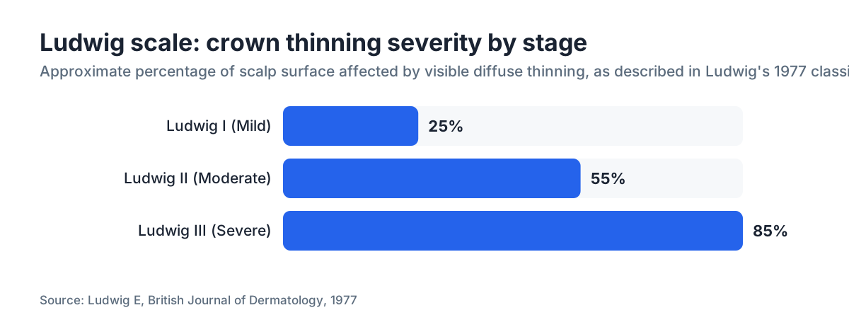
The Ludwig scale has three stages:
Ludwig I (Mild): Noticeable thinning on the crown and top of the scalp, but a band of denser hair remains near the frontal hairline. The part line looks wider than it used to. Most women notice it first when they look down at a part in bright light or fluorescent bathroom lighting.
Ludwig II (Moderate): More pronounced diffuse thinning across the crown and top, with the scalp clearly visible through the hair. The frontal hairline is still mostly preserved, which is the hallmark difference from male-pattern loss.
Ludwig III (Severe): Near-complete loss of hair across the top of the scalp, leaving only a thin rim of hair at the sides and back. This stage is less common and often has contributing factors beyond genetics alone, such as prolonged androgen excess or a long-untreated hormonal condition.
The one thing all three stages share is the preserved frontal hairline. That is almost the defining visual signature of female androgenetic alopecia, and it is the reason the Norwood scale, which tracks frontal recession, simply does not map onto it.
The Ludwig scale is described in the American Academy of Dermatology's clinical resources on hair loss and remains a standard teaching tool in dermatology training programs [3].
Ludwig vs. Norwood: what are the actual differences?
| Feature | Norwood Scale (male) | Ludwig Scale (female) |
|---|---|---|
| Number of stages | 7 main + variants | 3 |
| Pattern | Temple recession, crown thinning, merge | Diffuse crown thinning, hairline preserved |
| Frontal hairline | Recedes significantly | Mostly retained |
| Underlying driver | DHT acting on androgen-sensitive follicles | Same mechanism, different distribution |
| Published | 1975 (Hamilton 1951) | 1977 |
| Primary clinical use | Men with androgenetic alopecia | Women with androgenetic alopecia |
| Used for treatment dosing? | Sometimes for transplant planning | Less so; severity guides treatment more |
The difference is more than cosmetic. Male and female androgenetic alopecia share the same root mechanism, DHT binding to androgen receptors in genetically susceptible follicles and causing miniaturization [4]. But the distribution of sensitive follicles differs by sex. In men, the frontal and crown follicles are most sensitive. In women, the crown follicles are sensitive but the frontal hairline follicles are often somewhat protected, likely because of estrogen's partial buffering effect on androgen signaling at those follicles [5].
That biology is what produces the diffuse, top-of-scalp pattern Ludwig described, versus the progressive frontal recession Norwood maps. Mix up the tools and you get a misdiagnosis, which matters because treatment choices differ.
For anyone dealing with DHT blocker options or wondering about finasteride for female hair loss, staging the loss correctly is the foundation of having a productive conversation with a dermatologist.
Is there a newer or better scale for women's hair loss?
Yes. The Sinclair scale, published in 2004, has been gaining traction as a simpler, more practical alternative for women in clinical settings [6]. It uses five stages defined by the width of the midline part, which makes it easier to track over time with a ruler or a photograph.
Sinclair stages:
- Stage 1: Normal part width
- Stage 2: Part width widening
- Stage 3: Wide part, thinning visible across top
- Stage 4: Diffuse thinning, scalp clearly visible
- Stage 5: Advanced loss resembling Ludwig III
The part-width method has one real advantage: it gives patients a concrete thing to measure and photograph at home. The Ludwig scale requires some clinical judgment about the overall density pattern, which is harder for a patient to self-assess consistently.
Some dermatologists use the Savin scale, which is similar to Ludwig but adds a category for overall density and a separate scale for frontal thinning. A 2020 review in the Journal of the American Academy of Dermatology noted that no single scale has been universally adopted for women's hair loss research, partly because female androgenetic alopecia often overlaps with telogen effluvium and other shedding conditions, making clean staging harder [7].
Here is the short version. Ludwig is the most historically established. Sinclair is the most patient-friendly. A good dermatologist may use either, or both.
How do doctors use these scales to choose a treatment?
The scale tells a doctor where you are. It does not tell them why you are there. That distinction matters enormously.
A woman presenting at Ludwig II could have pure androgenetic alopecia, or a thyroid disorder, or chronic iron deficiency causing overlapping telogen effluvium, or a combination of all three. The scale points at the pattern; blood work points at the cause. Treatment before diagnosis is a common and expensive mistake.
Once the cause is reasonably clear, staging does influence treatment intensity:
Ludwig I: Often manageable with topical minoxidil 2% or 5% (both FDA-approved for women) [8]. At this stage, early intervention gives the best results because the follicles are miniaturizing but not yet dead.
Ludwig II: Topical minoxidil is still first-line. Spironolactone (an androgen blocker commonly used off-label for female pattern hair loss), low-level laser therapy, and platelet-rich plasma injections are often added. Oral minoxidil at low doses, typically 0.5 to 2.5 mg, is increasingly used off-label at this stage. Read more about oral minoxidil to understand how it differs from topical.
Ludwig III: The follicles across the crown may be permanently lost. A hair transplant becomes a serious conversation at this stage, though transplant surgeons are selective about female candidates because the donor area must be healthy and the diffuse nature of female loss can make transplanted follicles vulnerable.
Finasteride (1 mg daily) is FDA-approved for men but used off-label in postmenopausal women for female pattern hair loss. It is generally avoided in premenopausal women without reliable contraception because of teratogenicity risk. A dermatologist has to make that call individually.
The overlap with other conditions is also why hair loss supplements marketed heavily at women deserve skepticism: a supplement cannot fix androgenetic alopecia, though correcting a true nutritional deficiency (iron, biotin, zinc) can improve overall shedding.
Can women have the Norwood pattern of hair loss?
Rarely, yes. Some women, particularly those with polycystic ovary syndrome (PCOS), congenital adrenal hyperplasia, or androgen-secreting tumors, produce substantially elevated androgens and can develop hair loss patterns that look more like male pattern loss, including some degree of frontal recession.
There is also a genetic variant: a small subset of women with androgenetic alopecia do show Hamilton-Norwood type recession rather than Ludwig-type diffuse loss. This is uncommon enough that researchers have proposed a separate classification for it, sometimes called "female-pattern male hair loss" or Hamilton-type FPHL.
If a woman is losing hair at a receding hairline rather than across the crown, that is worth investigating for androgen excess rather than assuming it is standard female pattern loss. An endocrinologist or a dermatologist with a hormone focus is the right specialist. You can read more about receding hairline patterns to compare.
For women with PCOS-related hair loss, treatment may differ meaningfully from standard FPHL treatment, because managing the underlying hormonal condition is often the first-order intervention.
How do you figure out your own Ludwig stage?
You do not need a clinic to get a rough sense of where you are. Here is what to look at:
The part test. Part your hair straight down the middle under bright, direct light. Look at the width of the exposed scalp and compare it to an old photo. A progressively widening part on the crown side is the most reliable self-observable sign of Ludwig-pattern loss.
The density test. Pull a section of crown hair into a ponytail. Compare the circumference of that ponytail to photos from 5 or 10 years ago. A meaningful reduction in circumference signals significant diffuse thinning.
The hairline check. Is your frontal hairline roughly where it always was? If yes, that supports Ludwig-pattern loss. If you're seeing temple recession or a noticeably higher forehead, see a dermatologist sooner rather than later to rule out androgen excess.
Self-staging has limits. A significant percentage of women who think they have androgenetic alopecia turn out to have telogen effluvium, or both at once. A dermatologist uses dermoscopy (a magnified examination of the scalp) to assess follicle miniaturization directly, which is more definitive than any staging scale alone.
MyHairline's free AI hair scan at /scan can give you a starting visual assessment using your own photos, which is a practical first step before deciding whether a dermatologist visit is warranted.
Does hair loss progress through Ludwig stages over time?
For most women with androgenetic alopecia, yes, the loss is progressive. But the pace varies enormously and is hard to predict for any individual.
A study following women with androgenetic alopecia over several years found that a significant proportion remained stable at Ludwig I for extended periods, while a smaller subset progressed more rapidly [9]. The research could not reliably predict which women would progress quickly.
Factors that appear to influence progression speed:
- Onset age. Women who first notice thinning in their 20s or 30s tend to have more total loss by midlife than women whose loss starts after menopause, though this is not universal.
- Androgen levels. Higher circulating androgens, even within the normal range, are associated with faster progression.
- Treatment. Minoxidil, the only FDA-approved topical treatment for female androgenetic alopecia, does not reverse miniaturization but can slow or halt it in a meaningful percentage of users [8].
- Menopause. The drop in estrogen at menopause removes some of the hormonal protection against androgen effects, and many women notice accelerated thinning in the perimenopausal and early postmenopausal years.
The practical implication: earlier intervention is better. Holding Ludwig I for years is a more winnable goal than recovering from Ludwig III.
What treatments are FDA-approved specifically for female hair loss?
This is a short list, and it's worth being precise about it.
The FDA has approved topical minoxidil 2% solution for women (approved 1991) and topical minoxidil 5% foam for women (approved 2014) for the treatment of female androgenetic alopecia [8]. The label indication is specifically androgenetic alopecia, the Ludwig-pattern loss described above.
Nothing else carries an FDA approval specifically for female pattern hair loss as of 2025. That does not mean nothing else works. It means the formal approval process has not been completed for other agents.
Commonly used off-label options for women include:
- Spironolactone 25 to 200 mg daily (oral androgen blocker, very common in dermatology practice)
- Finasteride 1 to 2.5 mg daily (primarily in postmenopausal women)
- Low-level laser therapy devices (FDA-cleared for safety, not approved as a drug)
- Platelet-rich plasma injections (evidence is mixed; no FDA drug approval)
- Oral minoxidil 0.5 to 2.5 mg daily (off-label, growing evidence base)
For the minoxidil side effects that women should know about before starting treatment, that article covers the specifics.
Combining minoxidil with an androgen blocker is common practice for women who have clear androgenetic alopecia and tolerate the medications, similar to how finasteride and minoxidil are often combined in men.
Is the Ludwig scale used for hair transplant planning in women?
Hair transplants in women are more complex than in men, and the Ludwig scale plays a supporting role in planning rather than the central one.
For men, the Norwood stage is often the starting point for a transplant surgeon's assessment, because the predictable pattern helps identify where grafts go and how many are needed. For women, the diffuse nature of Ludwig-type loss creates a real problem: the donor area (the back and sides of the scalp) may itself be affected by diffuse thinning, meaning the "permanent" zone of hair that makes transplantation durable is less reliable [10].
Transplant surgeons typically require women to be at Ludwig II or Ludwig III, have a clearly identified cause for their loss (androgenetic alopecia confirmed, more than shed), show stable loss for at least a year, and have an adequate donor density. The Ludwig stage tells the surgeon about the recipient area. Dermoscopic assessment of the donor area tells them whether the surgery is viable.
Women with telogen effluvium or other diffuse shedding conditions are generally poor transplant candidates regardless of Ludwig stage, because the underlying shedding process can affect transplanted grafts.
Read more about hair transplant criteria if you're at a stage where that conversation feels relevant.
Are there conditions that mimic Ludwig-pattern hair loss?
Several. This is one of the more under-discussed problems in female hair loss: women correctly identify that their crown is thinning, get assigned a Ludwig stage, and start minoxidil, without anyone identifying the actual driver.
Telogen effluvium (TE). Diffuse shedding triggered by stress, illness, surgery, childbirth, or dramatic dietary restriction. It causes widespread thinning across the crown and can look like Ludwig I or II. The key difference: TE is often reversible once the trigger resolves. Androgenetic alopecia is not. The two can co-exist, which makes the picture messier.
Thyroid disease. Both hypothyroidism and hyperthyroidism cause diffuse hair loss. A TSH blood test is one of the first things any responsible doctor orders for a woman presenting with hair loss.
Iron deficiency. Serum ferritin levels below roughly 30 to 40 ng/mL are associated with hair shedding in some studies, though the cutoff is debated. This is correctable.
Scalp conditions. Frontal fibrosing alopecia (a scarring alopecia) can cause a different pattern but is sometimes initially mistaken for androgenetic alopecia.
This is why a diagnosis before treatment matters, and why no AI tool or staging scale replaces a dermatologist with a dermoscope and a blood panel. The scale tells you the pattern. It does not tell you why the pattern is there.
Sources
- Norwood OT. Male pattern baldness: classification and incidence. Southern Medical Journal, 1975.
- Ludwig E. Classification of the types of androgenetic alopecia (common baldness) occurring in the female sex. British Journal of Dermatology, 1977.
- American Academy of Dermatology Association – Hair loss types: Androgenetic alopecia
- Vary JC. Selected Disorders of Skin Appendages. Medical Clinics of North America, 2015 (PubMed).
- Blume-Peytavi U et al. S1 guideline for diagnostic evaluation in androgenetic alopecia in men, women and adolescents. British Journal of Dermatology, 2011.
- Sinclair R et al. A new scale for measuring female pattern hair loss. British Journal of Dermatology, 2004.
- Vañó-Galván S et al. Female pattern hair loss. Journal of the American Academy of Dermatology, 2020.
- FDA – Minoxidil topical solution and foam labeling
- Olsen EA. Female pattern hair loss and natural history. Journal of the American Academy of Dermatology, referenced female pattern progression data.
- Bernstein RM, Rassman WR. Follicular transplantation: patient evaluation and surgical planning. Dermatologic Surgery, 1997.
- American Academy of Dermatology Association – Diagnosing and treating hair loss
- Rossi A et al. Minoxidil use in dermatology: side effects and recent patents. Recent Patents on Inflammation and Allergy Drug Discovery, 2012.