

TL;DR: Telogen effluvium causes diffuse shedding across the whole scalp rather than patchy bald spots. You typically lose 300 to 500 hairs a day instead of the normal 50 to 100, see thinning at the part line and temples, and notice club-root hairs on your pillow. Regrowth usually starts within 3 to 6 months once the trigger is removed.
What does telogen effluvium actually look like?
The first thing most people notice is the shower drain. Where you used to see a few loose hairs, there's now a clump. Then it's the pillow, the hairbrush, the bathroom floor. That's the visual signature of telogen effluvium: shedding that feels dramatic and sudden, spread evenly across the whole scalp rather than piled into one spot.
Male-pattern baldness starts at the temples or crown and leaves those areas noticeably thinner. Telogen effluvium thins everything at once. The part line gets wider. The ponytail gets thinner. In bright light or against a dark background you can see scalp where you couldn't before. But you won't usually see a completely bare patch.
The hairs you're losing have a distinctive look too. Each one has a small white or pale bulb at the root end. That's the club hair, the follicle's way of packaging a strand for release at the end of its resting (telogen) phase. Seeing that bulb under a fingernail or on a white pillowcase confirms the hair was shed normally rather than snapped from damage.
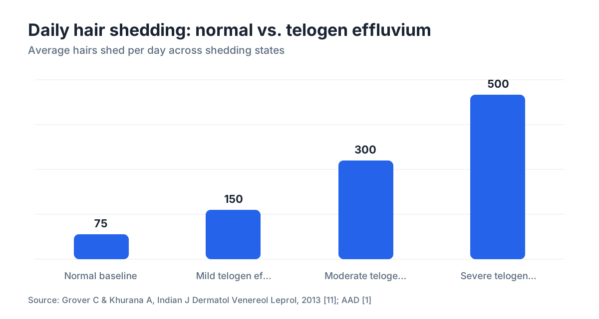
Normal daily hair loss runs about 50 to 100 strands, according to the American Academy of Dermatology [1]. In active telogen effluvium that number climbs to 300 or more. Some patients in clinical studies reported losing 400 to 500 strands daily during peak shedding. That jump is big enough to see with your own eyes when you count what's on the brush.
What do telogen effluvium hairs look like up close?
Pick one of the shed hairs and hold it near a light. The root end should have a small, rounded, slightly translucent bulb attached, sometimes white, sometimes pale yellow. That club root means the hair completed its growth cycle normally before being shed. It's different from a hair that tapers to a fine point at the root, which suggests it was pulled prematurely from an active (anagen) follicle.
The club hair is the single most useful close-up image for self-diagnosis. If nearly all your shed hairs have that club, you're seeing classic telogen effluvium. If many hairs taper to a thin tip or show a dark, gel-like root sheath, something else may be going on, and a dermatologist's trichoscopy will tell you more.
The hair shaft itself usually looks healthy in telogen effluvium. No splitting along the length, no obvious miniaturization (the progressive thinning of shaft diameter you see in androgenetic alopecia). The strand is the same width from root to tip. That's reassuring: the follicle is intact and capable of growing another hair.
A trichoscopy image, the kind a dermatologist takes with a handheld dermoscope or video system, shows empty follicular openings (the follicle shed the hair but hasn't grown a replacement yet), occasional short regrowing hairs, and normal density of follicular units in areas that haven't started regrowing [2]. You won't see the miniaturized follicles or "follicular dropout" that mark advanced androgenetic alopecia.
How does the scalp look with telogen effluvium vs. other types of hair loss?
Treatment splits completely depending on which condition you actually have, so this comparison earns its keep.
| Feature | Telogen Effluvium | Androgenetic Alopecia | Alopecia Areata |
|---|---|---|---|
| Pattern | Diffuse, whole scalp | Patterned (temples, crown) | Round or oval patches |
| Scalp appearance | Normal, no inflammation | Normal or visible scalp through thin hair | Smooth, bald patch, possible exclamation-point hairs at edges |
| Shed hair root | Club (white bulb) | Club + miniaturized shaft | Tapered or exclamation-point |
| Scalp inflammation | Absent | Absent | Sometimes slight redness at patch edges |
| Timeline | Acute shed 2-4 months post-trigger | Gradual over years | Days to weeks |
| Recovery | Usually full if trigger removed | Requires ongoing treatment | Variable; may recur |
In scalp photographs, telogen effluvium shows a wider part line and thinner density all over, but the scalp skin itself looks normal. No redness, no scaling, no follicular scarring. That's one of the clearest visual clues: lots of hair on your pillow, healthy-looking scalp underneath.
Alopecia areata leaves a distinct smooth bald patch. The skin inside the patch sometimes looks slightly shiny. At the edges you might see "exclamation point" hairs, short strands narrower at the base than the tip, a hallmark of that condition [10]. You'll never see that in telogen effluvium.
Androgenetic alopecia builds over years. Early-stage images show miniaturized hairs (fine, short, lighter-colored) mixed in with normal terminal hairs, densest at the temples and crown. Diffuse thinning without that zonal concentration points back to telogen effluvium. The two can coexist, which makes a dermatology assessment genuinely useful if you're unsure. Learn more about what causes hair loss to understand which other conditions might look similar.
What does the part line look like in telogen effluvium?
The part line is the easiest place to photograph at home and the most telling. In healthy hair, parting reveals a narrow strip of scalp. In moderate telogen effluvium that strip widens noticeably, sometimes doubling or tripling in visible width, because the hairs that once flanked the part have shed.
A comparison you can do yourself: part your hair the same way in a well-lit room (bathroom light above you works) and photograph from directly overhead. Compare to a photo from six months earlier. If the strip of visible scalp is wider and the individual hairs framing it look finer, that lines up with diffuse telogen shedding.
In androgenetic alopecia (female pattern specifically) the part also widens, but the shape differs. It forms a "Christmas tree": wider at the front of the scalp and narrowing toward the crown, with the frontal hairline preserved. Telogen effluvium widens the part more uniformly, without that front-heavy concentration [3].
This difference shows up in side-by-side clinical photographs used in dermatology training. The frontal hairline in telogen effluvium stays relatively intact. Losing density at the part while your hairline edge holds is a visual reassurance that you're not dealing with a receding hairline from androgenetic alopecia. If your hairline is also moving back, read about receding hairlines separately, because the management differs.
What do regrowth hairs look like after telogen effluvium?
Regrowth is the visual sign people most want to see and most often miss. It starts around 3 to 6 months after the trigger resolves [1], and the first hairs to emerge are short, fine, and often slightly lighter in color than your mature hair. They look like a fringe of baby hairs along the hairline and part.
In close-up photographs or trichoscopy images, regrowing hairs appear as short upright strands, sometimes less than a centimeter long, sticking up from the scalp surface. Under dermoscopy they show as thin, tapered shafts emerging from follicular openings that were recently empty. This "forest of short hairs" alongside longer mature hairs is a classic regrowth image and a strong positive sign.
The regrowth phase can itself feel alarming. Your hair looks uneven, the new short hairs create a different texture, and some people mistake the two-length appearance for ongoing thinning. It isn't. If you see those short hairs at the hairline and temples, the follicles are working.
Full cosmetic recovery usually takes 6 to 12 months from the start of regrowth, depending on how long your hair was before the shed. Longer hair takes more time because each centimeter of length takes roughly a month to grow, at the average rate of about 1.25 cm per month [4]. Photographing the part line every four weeks gives you a concrete record of whether you're progressing.
How much shedding is actually visible, and how do you document it?
The clinical threshold dermatologists use for a meaningful shed is more than 100 hairs per day, confirmed by a standardized 60-second hair-count or a trichogram [2]. For everyday purposes, though, the visual accumulation is what most people notice first.
A practical home method: after not washing for 24 hours, shampoo normally and collect every hair from the shower drain and your hands in a white bowl or on a white towel. Count them, or photograph them against a white background for comparison. Repeat weekly and you get a visual trend. A cluster of 200 or more hairs after one wash is worth noting; a cluster that shrinks over successive weeks suggests the acute shed is winding down.
Photography tips that actually capture what's happening:
- Use a well-lit room with overhead light, not backlighting.
- Part your hair in the same place every time, photographed from directly above.
- Include a ruler or credit card in the frame for scale when you photograph shed hairs.
- Wet hair photographs differently than dry hair. Wet hair clumps and can look thinner or thicker depending on technique. Stick to one method.
If you want an AI-assisted read on what your photos show, MyHairline's free scan tool at myhairline.ai/scan can compare your scalp density and part-line width against reference data and flag patterns worth discussing with a dermatologist. It doesn't replace a clinical exam. It gives you a structured starting point.
Can you see telogen effluvium in blood tests or trichoscopy, more than photos?
Scalp photographs tell part of the story. Trichoscopy (dermoscopy applied to the scalp) adds another layer. Under magnification a dermatologist looks at the ratio of terminal to vellus hairs, the number of empty follicular openings, and the density of follicular units per square centimeter. In acute telogen effluvium, follicular units are present but empty or growing short anagen hairs, with no significant miniaturization [2].
The trichogram is a different tool. It involves plucking 50 to 60 hairs from a specific scalp region and examining their roots under a microscope. A normal telogen percentage is around 10 to 15 percent. In active telogen effluvium it rises above 25 percent, sometimes reaching 50 percent or higher in severe cases [5]. That percentage is a definitive diagnostic image, just one you need a microscope to see.
Blood tests don't produce images of hair loss, but they identify the triggers. A standard workup includes ferritin (low iron is one of the most common correctable causes), thyroid-stimulating hormone, complete blood count, and sometimes zinc and vitamin D [12]. The American Academy of Dermatology recommends these labs for patients presenting with diffuse hair shedding [1].
Put it together and the picture is strong: diffuse shedding, a club-root-dominant trichogram, a widened part line, and a relevant trigger in the history (surgery, illness, crash diet, childbirth six months prior). Most dermatologists are comfortable confirming the diagnosis on that combination without a scalp biopsy, though biopsy remains the gold standard when the diagnosis is genuinely ambiguous.
What triggers cause this pattern and show up in patient timelines?
The visual shed typically peaks about 2 to 4 months after the triggering event [3]. That delay is why so many people feel confused. The dramatic hair loss appears long after they felt sick, gave birth, or ended a crash diet, so they don't connect the two.
Common documented triggers:
Physical stress: major surgery, high fever, severe illness (COVID-19 has been linked to significant telogen effluvium in several documented case series). Childbirth is the classic cause of postpartum hair loss, typically appearing 2 to 4 months after delivery.
Nutritional deficiency: iron deficiency (especially low ferritin, even without frank anemia), rapid weight loss, protein restriction. Studies have found that ferritin below 30 ng/mL is frequently present in women with diffuse hair shedding [5].
Medications: certain blood pressure drugs (beta-blockers), retinoids, anticoagulants, and others can push follicles into telogen. The FDA label for isotretinoin, for example, lists hair loss as a reported adverse effect [6].
Psychological stress: the evidence here is less clean. Extreme psychological stress probably can trigger a telogen shift, but the effect is harder to quantify and the studies are smaller. For a full rundown of every cause, the what causes hair loss guide is a good companion read.
Chronically elevated stress or ongoing nutritional problems can cause a persistent (chronic) form lasting more than six months, sometimes years [9]. The visual presentation looks like acute telogen effluvium but keeps going because the trigger hasn't resolved. If you're months into shedding with no sign of improvement, tracking down and eliminating the underlying cause matters more than any topical treatment.
Does minoxidil change what telogen effluvium looks like?
Minoxidil is FDA-approved for androgenetic alopecia, not telogen effluvium specifically [7]. Some dermatologists use it off-label in prolonged cases anyway, because it can shorten the resting phase and push follicles back into active growth.
Here's the visual catch. Starting minoxidil often causes an initial shedding surge in the first 2 to 6 weeks. It happens because minoxidil shifts follicles out of telogen, and the old club hairs fall out before the new anagen hairs emerge. If you're already in telogen effluvium and you add minoxidil, that early shed can look scary, more hairs in the shower than before. It's usually temporary, but it's worth knowing about before you start.
The regrowth hairs that appear after minoxidil use look like the natural regrowth described earlier: short, fine, upright hairs along the part and hairline. The difference is they may show up a few weeks sooner than they would have without treatment. Read the full breakdown of minoxidil for men and minoxidil side effects if you're weighing whether to add it.
Finasteride is not a standard treatment for telogen effluvium. It targets DHT-driven miniaturization in androgenetic alopecia. If both conditions are present (which happens), finasteride and minoxidil together address the androgenetic component while the telogen effluvium resolves on its own. A dermatologist can look at your scalp images and blood work to sort out whether you're dealing with one condition or both.
When should the images you see make you call a dermatologist?
Most cases of telogen effluvium resolve on their own within 6 to 12 months once the trigger is gone. You probably don't need urgent care if the shed is acute, you can name a likely trigger from roughly 2 to 4 months before the shedding started, and your scalp skin looks normal in photos.
See a dermatologist if:
Shedding has continued for more than 6 months with no visible regrowth phase. That duration pushes toward chronic telogen effluvium or points to an ongoing underlying cause nobody has found yet [9].
Your photos show smooth, defined bald patches rather than diffuse thinning. Patches suggest alopecia areata or other autoimmune conditions that need specific treatment.
You see scalp redness, scaling, or follicular pus in photographs. Those signs point to inflammatory scalp conditions (seborrheic dermatitis, folliculitis, lichen planopilaris) that need different management entirely.
You notice a receding hairline at the temples alongside the diffuse shed. That combination may mean androgenetic alopecia is present at the same time, and the evidence base for treating it is strong: FDA-approved minoxidil for men and finasteride both have published clinical trial support [7][8].
You've had blood work done, everything looks normal, and the shedding continues anyway. A scalp biopsy is the most definitive diagnostic image when clinical assessment hasn't given you a clear answer. It can distinguish telogen effluvium, early androgenetic alopecia, and early scarring alopecias before the visual surface changes become obvious. MyHairline's free AI scan can help you document the pattern before your appointment and give your dermatologist a baseline image to compare against at follow-up visits.
What's a realistic visual timeline from peak shed to full recovery?
Laying this out concretely helps, because the waiting is the hardest part.
Month 0: Trigger event (surgery, illness, delivery, crash diet, major stress).
Months 1 to 2: Follicles shift into telogen. No visible change yet, because the hairs are still attached; they're just not actively growing.
Months 2 to 4: Peak shedding. This is when the drain, the pillow, and the brush become alarming. The part line widens visibly. A photograph now versus four months earlier shows the difference clearly.
Months 3 to 6: Shedding slows as new anagen hairs begin emerging. Short, fine regrowth hairs appear at the hairline and part. The texture changes: longer hairs alongside new short ones.
Months 6 to 12: Density returns. The part line narrows toward its old width. Ponytail circumference recovers. Photographs every four weeks show a clear trend toward baseline.
Month 12 and beyond: Most patients with a single acute trigger report full or near-full cosmetic recovery by 12 months [3]. Patients with chronic telogen effluvium or an unresolved trigger take longer, and some permanent thinning is possible if the chronic form persists for years, though this is uncommon.
The visual journey is genuinely slow. The gap between "shedding stops" and "hair looks normal again" is months, not weeks, because every new hair has to grow from zero length to cosmetically visible length at roughly 1.25 cm per month [4]. That timeline is frustrating but predictable, and predictability is its own kind of reassurance.
Sources
- American Academy of Dermatology (AAD), Hair loss types and hair shedding guidance
- Rudnicka L et al., Atlas of Trichoscopy, Springer (2012), trichoscopy findings in telogen effluvium
- Malkud S. Telogen Effluvium: A Review. Journal of Clinical and Diagnostic Research. 2015;9(9):WE01-WE03.
- Trüeb RM. Physiology and Pathophysiology of Hair Growth. Skin Pharmacology and Physiology. 2018.
- Trost LB, Bergfeld WF, Calogeras E. The diagnosis and treatment of iron deficiency and its potential relationship to hair loss. J Am Acad Dermatol. 2006;54(5):824-844.
- FDA, Isotretinoin (Accutane) prescribing information / label
- FDA, Minoxidil topical solution approved labeling (Rogaine NDA)
- Finasteride (Propecia) FDA approved labeling, Merck
- Hughes EC, Saleh D. Telogen Effluvium. StatPearls, NCBI Bookshelf. National Library of Medicine.
- NIH National Institute of Arthritis and Musculoskeletal and Skin Diseases (NIAMS), Alopecia Areata
- Grover C, Khurana A. Telogen effluvium. Indian J Dermatol Venereol Leprol. 2013;79(5):591-603.
- Almohanna HM et al. The Role of Vitamins and Minerals in Hair Loss: A Review. Dermatol Ther (Heidelb). 2019;9(1):51-70.