

TL;DR: Miniaturized hairs are thin, short, pale strands that grow next to normal hairs in areas hit by androgenetic alopecia. Find them at home by parting dry hair in bright light and using a 10x magnifier or a phone macro lens. Catching them early matters because minoxidil and finasteride work best before the follicle dies off completely.
What are miniaturized hairs, exactly?
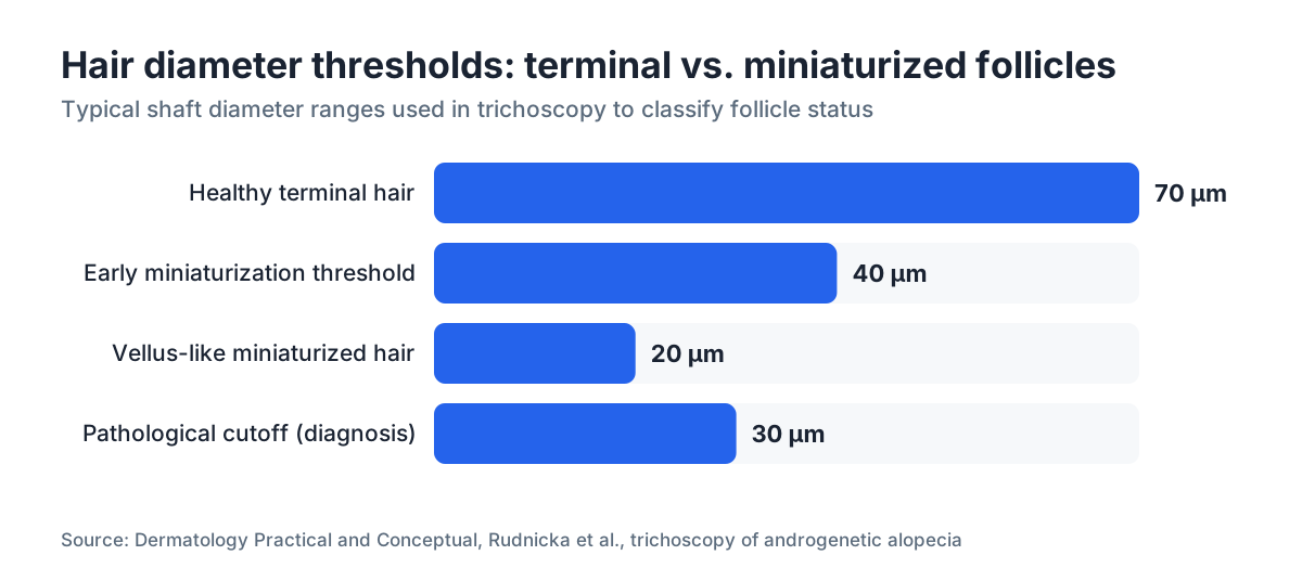
A healthy terminal hair is roughly 60 to 80 micrometers thick and pigmented the whole way down the shaft. A miniaturized hair, sometimes called a vellus-like hair, has shrunk to a fraction of that, often under 30 micrometers, and lost most of its color. It reads as fine, nearly colorless fuzz instead of a real strand. [1]
The process is called follicular miniaturization, and it is the physical mechanism behind androgenetic alopecia, the most common form of hair loss in men and women. Dihydrotestosterone (DHT) binds to receptors in genetically susceptible follicles and shortens the anagen (growth) phase with each cycle. The follicle shrinks over years, producing thinner, shorter, lighter hairs until it stops producing a visible hair at all. [2]
Timing is everything here. Once a follicle has fully atrophied and been replaced by fibrotic tissue, minoxidil and finasteride cannot bring it back. The window for treatment is while miniaturization is happening, not after. Finding these hairs at home gives you an early read on whether that window is still open. There is more on what causes hair loss and where DHT fits in.
Miniaturized hairs are not shed hairs. A shed hair has a white bulb at the root. A miniaturized hair is still attached to the scalp and still growing, just producing a thin, short strand instead of a normal one.
How do miniaturized hairs look and feel different from normal hairs?
The fastest way to see the difference is to lay two hairs side by side on white paper.
A terminal hair on paper is clearly visible, maybe 2 to 5 centimeters long if pulled mid-cycle, with obvious color. A miniaturized hair from the same area is nearly invisible against the paper. It may be under a centimeter long even after the same growing time, because anagen has been cut short. Zoom your phone camera to its macro limit and you can watch the shaft taper to almost nothing, missing the smooth cylindrical profile of a healthy hair.
On the scalp, miniaturized hairs feel like soft fuzz when you drag a fingertip slowly across the surface. Normal hairs push back. Miniaturized hairs barely register.
Color is a reliable tell. A miniaturizing follicle makes less pigment, so the hairs look lighter than the rest of your hair even if your hair is naturally dark. This is why thinning crowns often look pale in photos before the skin itself shows through.
Hair diameter variation across a single patch is the most diagnostic feature dermatologists look for. Trichoscopy studies show that a follicular unit holding hairs of very different diameters, more than 20 percent variation, is a hallmark of androgenetic alopecia. [3] That same variation shows up at home with the right setup.
What equipment do you need to find miniaturized hairs at home?
You do not need a clinic-grade trichoscope. At home the goal is enough magnification and light to see shaft diameter differences. Here is what actually works, ranked by how well it works.
| Tool | Magnification | Cost (approx.) | Honest assessment |
|---|---|---|---|
| Dermatoscope (handheld, polarized) | 10x | $30, $80 | Best home option; built for scalp and skin [4] |
| Digital USB microscope | 50x, 200x | $25, $60 | Great detail but awkward on your own scalp |
| 10x jeweler's loupe | 10x | $8, $20 | Works well with a second person helping |
| Macro lens clip for smartphone | 15x, 20x | $10, $25 | Convenient; quality varies by brand |
| Smartphone camera, pinch zoom, bright light | ~2x, 3x effective | $0 | Better than nothing; misses subtle cases |
| Naked eye in bright light | 1x | $0 | Catches obvious cases; misses early stages |
The single best low-cost upgrade is a handheld polarized dermatoscope. Polarized light cuts surface glare and makes fine shaft detail much clearer. Many are marketed for checking moles but work just as well on follicles.
Light matters as much as the lens. Direct sunlight or a high-CRI (color rendering index 90+) LED held close to the scalp is the difference between seeing nothing and seeing clearly. Bathroom ceiling light alone almost never cuts it.
Which areas of the scalp should you check first?
Androgenetic alopecia follows predictable patterns. In men it usually starts at the temples and crown (vertex). In women it shows as diffuse thinning along the central part, with the frontal hairline often spared. [5] So start your exam at those high-probability zones.
For men: check the crown first. Part the hair in a few directions under bright light and look for zones where hairs are noticeably finer and lighter than the surrounding hair. The temples are the second spot. Compare density and caliber at the very front edge against hair from the back of your head, which is largely DHT-resistant.
For women: part your hair straight down the middle from forehead to crown. The central part is the most sensitive early indicator. If the part looks wider than about 0.5 centimeters, or the hairs nearest the part are visibly finer than hairs further out, miniaturization is likely underway. A Christmas tree pattern, wide at the front and tapering back, is a recognized clinical sign of female pattern hair loss. [6]
For anyone: the occipital area (back of the head, above the nape) is mostly DHT-resistant and makes a good personal baseline. Hair there is your "normal" caliber. Judge anything you find on the crown or temples against it.
One practical habit: take comparison photos in the same lighting, same part, same distance, once every three months. Single-session looks are hard to read. Change over time is what tells you whether miniaturization is actively moving.
Step-by-step: how to do the self-examination
Step one: wash and fully dry your hair. Wet hair clumps and hides fine strands. Dry hair lets each shaft stand on its own.
Step two: set up your light. Sit by a window with direct daylight, or hold a bright LED torch at a low angle to the scalp. A low angle throws shadows that make fine hairs pop against the skin.
Step three: use a fine-tooth comb to part the hair in a system. Start at the center part and work outward in half-inch sections. Go slow.
Step four: look with your magnifier held close to the scalp. You want two things: hairs that are clearly thinner or shorter than their neighbors in the same follicular unit, and follicular units holding a mix of thick and thin hairs side by side.
Step five: pluck one suspect hair and one clearly normal hair from the back of your head. Lay both on a white index card in good light. Compare shaft diameter by eye. If the suspect hair is obviously thinner, you have found a miniaturized strand.
Step six: photograph what you find. Shoot the part line and crown from a fixed distance, about 12 inches straight above, every few months. That photo archive becomes your own longitudinal record, far more useful than any single snapshot.
Want a faster read? MyHairline's free AI scan can analyze your scalp photos for signs of miniaturization and density loss, useful alongside your own exam.
Step seven: note the location. Keep a phone note: date, which zones looked affected, rough subjective severity. Dermatologists find this history useful when you eventually see one.
How accurate is a home check compared to a dermatologist trichoscopy?
Honestly, a home check is less accurate. A clinical trichoscopy using a video dermatoscope at 20x to 70x with calibrated image software can measure follicular unit density, shaft diameter, and the terminal-to-vellus ratio with a precision no phone camera touches. [3]
But a home check is far from useless. It does two things well: catching obvious, real miniaturization early enough to get professional evaluation, and tracking change between appointments.
The biggest limitation is that you cannot measure your own follicular unit density reliably at home. Clinical diagnosis of androgenetic alopecia usually uses a terminal-to-vellus ratio below 4:1 as the threshold for pathological miniaturization. [3] You will not calculate that ratio with a loupe and a phone. What you can do is notice that some hairs look completely different from others in the same area, and that is enough to act on.
Another limit: telling miniaturization apart from telogen effluvium, which sheds hair without necessarily miniaturizing it, is genuinely hard at home. Telogen effluvium tends to drop normal-caliber hairs with white club roots, while androgenetic alopecia produces fine, short, attached hairs. There is overlap, and both can run at once.
If your home exam finds obvious miniaturization, especially if it is progressing in photos over three to six months, book a dermatology appointment rather than second-guessing yourself.
What does the research say about how early miniaturization can be detected?
Miniaturization starts well before the scalp looks thin. A 2000 study in the Journal of Investigative Dermatology found that follicular miniaturization can be confirmed histologically in areas that look clinically normal in men with androgenetic alopecia, meaning the process starts years before any cosmetic hit. [1]
Trichoscopy studies confirm diameter variation shows up early. Research published in Dermatology Practical and Conceptual reported peripilar signs and hair shaft heterogeneity in early-stage androgenetic alopecia at Norwood-Hamilton Stage II to III in men and Ludwig Stage I in women, stages most people would not yet call significant. [3]
The practical read: if you check regularly in your mid-to-late twenties or thirties and you have a family history of hair loss, you have a real shot at catching miniaturization early enough that finasteride, minoxidil, or the two together could slow or partly reverse it. The American Academy of Dermatology says treatment works best while follicles are still present and producing some hair, not after the follicle is gone. [5]
Nobody has good longitudinal data on how many people use home detection to catch loss early and then treat it successfully. That study has not been done. The mechanistic case for early detection and early treatment, though, is solid.
What should you do if you find miniaturized hairs?
Finding miniaturized hairs is not a diagnosis. It is a signal to act.
Start with a board-certified dermatologist or a trichologist. They can run a formal trichoscopy, take a scalp biopsy if needed, rule out other causes like scalp conditions or nutritional deficiencies, and confirm whether what you found is androgenetic alopecia.
If it is confirmed, the two treatments with the strongest evidence are topical or oral minoxidil and, for men, finasteride. The FDA approved topical minoxidil for men (5%) and women (2%) for androgenetic alopecia, and the labeling states it "has not been shown to regrow hair in most women" once follicles are dead, which is exactly why starting early matters. [7]
Finasteride, a DHT blocker, is FDA-approved for men with androgenetic alopecia at 1 mg daily. A 5-year trial showed 48 percent of men on finasteride had increased hair count at year 5, against 75 percent of the placebo group who kept losing hair. [8] There is more on finasteride and whether the evidence maps to your situation.
Some men and women run both together. The combination beats either alone in head-to-head comparisons, though the extra benefit has to be weighed against possible minoxidil side effects and finasteride's known effects profile. More on that at finasteride and minoxidil combined.
If miniaturization is very advanced and large areas are already bald, a hair transplant may be the realistic path, though transplants still need the miniaturization stabilized medically first, or the transplanted area will thin too.
Do not spend money on hair loss supplements as a first move. Most have no controlled trial evidence at anything near a meaningful effect size. Address the DHT-driven miniaturization directly first.
Can miniaturized hairs be reversed, or are they gone permanently?
They can be partly reversed if caught early. "Partly" is doing real work in that sentence.
A follicle still making a miniaturized hair still has functional papilla cells. Cut the DHT signal, or add minoxidil's vasodilatory and potassium channel effects, and some of those follicles return to producing a thicker, longer hair. This is reactivation rather than regrowth in the strict sense, because the follicle was never fully dead.
Finasteride trials show meaningful hair count gains over two years in men with mild to moderate loss, with regrowth concentrated at the vertex. [8] Minoxidil trials in women show the 5% formulation produced significantly more regrowth than the 2% in a 48-week randomized controlled trial. [9] Both treatments hit diminishing returns once follicles are fully miniaturized and fibrotic.
The honest answer: some miniaturized follicles can be pulled back toward normal, especially in the first few years, but a follicle that has been miniaturizing for a decade with no help is much less likely to respond. This is the whole reason early detection matters.
A DHT blocker is the most direct drug for slowing miniaturization in susceptible follicles. If you are a man in your twenties or thirties seeing early miniaturization and you have no contraindications, that conversation with a dermatologist is worth having soon rather than later.
Are there any common mistakes that make home detection harder?
Several, and they matter.
Checking wet hair is the biggest one. Wet hair bunches and hides fine individual shafts completely. Always examine dry hair.
Using bathroom lighting is the next most common. Ceiling fixtures throw flat, even light that washes out the detail you need. Use directional light at a low angle to the scalp.
Comparing yourself to old photos shot in different lighting or at a different distance is worse than useless. It creates false signals both ways. If you track change with photos, standardize the setup: same spot, same time of day, same distance, same part.
Ignoring the back of your head as a baseline is a quiet mistake. Without a reference from your DHT-resistant occipital region, you cannot calibrate what "normal" shaft diameter looks like for you. Your hair might be naturally fine or naturally coarse. Comparing to yourself beats a generic standard.
Panicking over normal variation is real too. Every scalp has some caliber variation. Not every thin hair is miniaturized. One thin hair in an otherwise dense, thick area is much less worrying than a cluster of thin hairs in the crown surrounded by thinning neighbors.
Checking once and calling it settled is a mistake. Hair loss is slow. A single exam tells you little. Serial looks over three to six months tell you whether a pattern exists and whether it is moving.
When should you stop self-examining and see a doctor?
See a dermatologist if any of these are true.
You find obvious clusters of miniaturized hairs in the crown or along the part, especially if the area has changed noticeably in your photos over three to six months.
You are losing more than roughly 100 hairs a day on a consistent basis. The American Academy of Dermatology says shedding 50 to 100 hairs daily is normal, and sustained shedding beyond that warrants evaluation. [5]
You have other symptoms with the hair changes: scalp itching, flaking, redness, pain, or patchy loss rather than diffuse thinning. Patchy loss can point to alopecia areata, an autoimmune condition that looks and behaves very differently from androgenetic alopecia and needs different treatment.
You are a woman under 40 with accelerating diffuse thinning. In women, pattern loss can be driven by hormonal conditions like polycystic ovary syndrome or thyroid dysfunction that deserve blood work before any topical treatment. [6]
You have a receding hairline that has moved back more than about a centimeter from where it was a year ago.
The bar for seeing a dermatologist about hair loss should be lower than most people set it. A trichoscopy takes about 15 minutes. Early diagnosis buys you more options. Waiting until the thinning is cosmetically obvious means you have already lost time you cannot get back.
Once you have finished your self-exam, if you want a second opinion on what your scalp photos show before booking, MyHairline's free AI scan can flag density and miniaturization patterns from your photos. It is not a medical diagnosis, but it helps you decide whether what you found is worth chasing.
Sources
- Journal of Investigative Dermatology, Whiting DA (2000), Diagnostic and predictive value of horizontal sections of scalp biopsy specimens in male pattern androgenetic alopecia
- American Academy of Dermatology, Hair loss types: Androgenetic alopecia
- Dermatology Practical and Conceptual, Rudnicka L et al., Trichoscopy of androgenetic alopecia
- National Center for Biotechnology Information (NIH), StatPearls: Dermatoscopy
- American Academy of Dermatology, Hair loss: Overview and self-care
- American Academy of Dermatology, Hair loss in women
- FDA, Minoxidil topical solution drug labeling (NDA 019501)
- New England Journal of Medicine, Kaufman KD et al. (1998), Finasteride in the treatment of men with androgenetic alopecia
- Journal of the American Academy of Dermatology, Lucky AW et al. (2004), A randomized, placebo-controlled trial of 5% and 2% topical minoxidil solutions in the treatment of female pattern hair loss
- Clinical Journal of Sport Medicine, van der Merwe J et al. (2009), Three weeks of creatine monohydrate supplementation affects dihydrotestosterone to testosterone ratio in college-aged rugby players
- National Institutes of Health, MedlinePlus: Hair loss