

TL;DR: Miniaturized hairs are full follicles that have shrunk below roughly 0.03 mm in diameter because of DHT exposure. Finding them in the frontal scalp or vertex before visible thinning starts is the clearest early sign of androgenetic alopecia (AGA). Dermatoscopy detects miniaturization as low as 10-20%, and treatment started at that stage consistently outperforms treatment started after obvious thinning.
What is hair miniaturization and why does it matter for AGA?
Hair miniaturization is a specific biological process, more than thinning. A follicle that is miniaturizing goes through progressively shorter anagen (growth) phases and produces a progressively thinner, shorter shaft until it eventually produces only a tiny vellus-like fiber, then nothing at all. The follicle is still alive during most of this process, which is why early intervention can actually reverse it.
In androgenetic alopecia, the trigger is dihydrotestosterone (DHT) binding to androgen receptors inside the dermal papilla of susceptible follicles. DHT shortens the anagen phase cycle by cycle. A follicle that once spent five years growing a thick terminal hair may, after several DHT-shortened cycles, spend only a few months growing a fine, lightly pigmented hair that barely clears the scalp surface [1].
This matters for treatment timing for one hard reason: once a follicle has been miniaturizing for many years and the dermal papilla shrinks below a critical size, minoxidil and finasteride cannot meaningfully regrow it. Several clinical trials show the best responders to finasteride are men who still have some miniaturized hairs remaining in the target zone, not completely bare follicles [2]. Catching miniaturization early, before you can see broad thinning in the mirror, is the window where medicine actually works.
The question most people have is: what does a miniaturized hair actually look like, and can I find them myself?
What does a miniaturized terminal hair look like compared to a normal one?
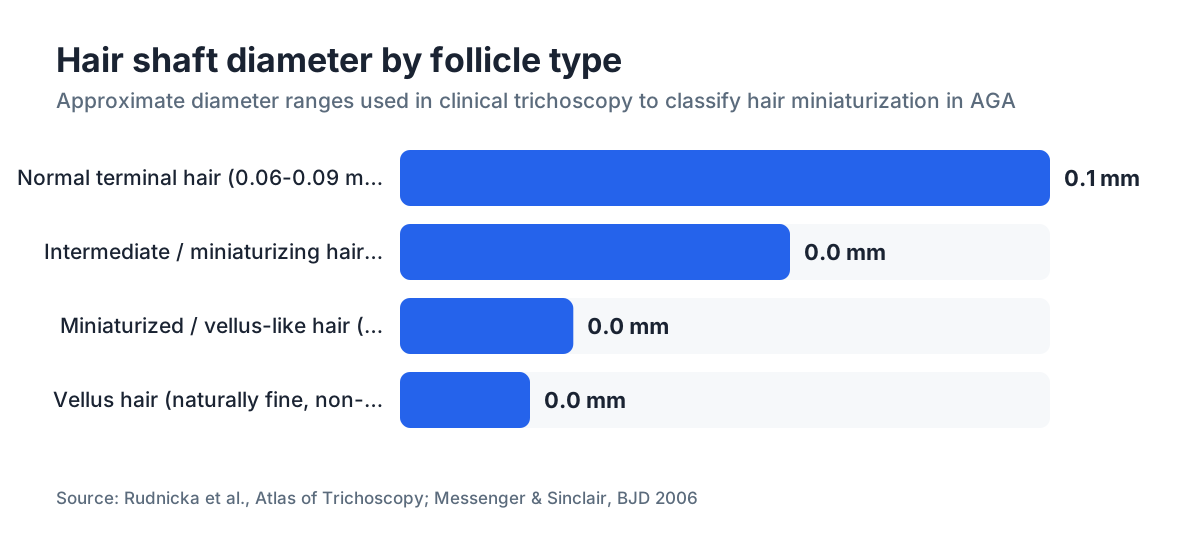
A normal terminal scalp hair has a shaft diameter between roughly 0.06 mm and 0.09 mm, visible to the naked eye as a thick, pigmented strand. A fully miniaturized hair, sometimes called an intermediate hair or vellus-like terminal hair in the dermatology literature, has a diameter under 0.03 mm and may look almost colorless, wispy, or nearly transparent in direct light [3].
The key distinguishing feature is the diameter-to-length ratio. Vellus hairs (the fine hairs on your forehead or cheek) are naturally small and short. Miniaturized terminal hairs are abnormal: they grew from a follicle that used to make thick hairs, and the shaft is disproportionately thin for its position on the scalp. If you pull one and look at it under a hand lens, the root end often appears thin and fragile compared to a normal club root.
Color is another clue. Melanin production in the hair shaft correlates with follicle size. As a follicle miniaturizes, it produces less melanin, so the same person's frontal hairs may look noticeably lighter or more translucent than their occipital hairs, which are DHT-resistant. This color gradient, frontal lighter than back and sides, is a real early sign and something a good dermatologist will comment on immediately.
Length also changes. Miniaturizing hairs do not reach their original full length because the shortened anagen phase stops growth earlier. So in the frontal zone you may notice short, wispy hairs that seem to never grow past a certain point while the rest of your hair grows normally. That is not breakage. That is a miniaturized follicle hitting the end of its shortened anagen.
| Feature | Normal terminal hair | Miniaturized hair |
|---|---|---|
| Shaft diameter | 0.06 to 0.09 mm | < 0.03 mm |
| Shaft color | Pigmented, matches rest | Lighter, translucent |
| Length potential | Full anagen 2-6 years | Shortened, may not pass 1-2 cm |
| Root under lens | Thick club | Thin, tapered or fragile |
| Location in AGA | Seen later in diffuse loss | Frontal/vertex, early sign |
Can you spot miniaturized hairs at home without special equipment?
Honestly, yes, partially, but with real limitations. The naked eye in normal room lighting will miss most early miniaturization. What you can do at home is set up conditions that make the subtlest hairs visible.
The single best DIY method is wet-hair examination in bright, direct light. Wash your hair and comb it flat without product. Stand under a bright LED bulb or go outside in direct sunlight. Part the hair in the areas classically affected by AGA: the frontal hairline (especially the temples and mid-frontal scalp), the vertex (crown), and the anterior mid-scalp. Look for hairs that are visibly finer and shorter than the surrounding hairs. Take a photo with your phone camera zoomed in, then compare to a photo taken six months later. The comparison over time is more informative than any single observation.
A $15-30 USB dermatoscope sold for skin examination will genuinely help. At 10x magnification you can see individual hair shaft diameters well enough to notice clear disparity between thick and thin hairs in the same zone. At 50x (the range many phone-attachment lenses reach) you can see the difference between a 0.06 mm and a 0.03 mm shaft plainly. You are not going to measure millimeters at home, but the visual contrast is obvious once you know what you are looking for.
What you cannot reliably do at home is separate miniaturized AGA hairs from the fine hairs caused by telogen effluvium or nutritional deficiency, because those conditions produce their own fine shedding. That distinction matters because the treatment is completely different. A dermatologist with a clinical dermatoscope and sometimes a scalp biopsy is the only person who can give you a confident answer.
Still, if you find a cluster of fine, short, light-colored hairs sitting in your frontal scalp alongside thicker hairs, especially if your father or grandfather had AGA, the probability is high that you are looking at early miniaturization. Starting with your what causes hair loss baseline and tracking it seriously is a legitimate first step.
How do dermatologists diagnose miniaturization: dermoscopy and hair analysis
The clinical gold standard for detecting miniaturization is trichoscopy, which is dermoscopy applied to the scalp and hair. A handheld dermatoscope at 10-20x magnification lets a dermatologist or trichologist directly see hair shaft thickness variation across a given area [3].
The key measurement is the hair diameter diversity index. A healthy scalp has relatively uniform hair shaft diameters in any given zone. In AGA, the same zone shows a mix of thick and thin hairs. Most published criteria define significant miniaturization as more than 20% of hairs in the affected zone showing a diameter less than half the average diameter for that person's scalp [4]. Some researchers use a simpler threshold: if more than 10% of hairs in the frontal or vertex zone appear clearly thinner than the occipital hairs, AGA is likely.
Beyond visual dermoscopy, there are two quantitative tools used in research settings and some specialty clinics. The first is the TrichoScan, a software system that photographs a small shaved area of scalp and automatically measures hair density, shaft diameter, and the ratio of terminal to vellus hairs [5]. The second is a manual phototrichogram, where a small area is shaved and photographed at two time points (usually 48-72 hours apart) to calculate anagen-to-telogen ratios and growth rates. Both methods are more precise than eyeballing but also more expensive and less widely available.
A scalp biopsy with horizontal sectioning is the definitive answer when the diagnosis is genuinely uncertain. Pathology can count the total follicular units per square centimeter, measure the terminal-to-vellus hair ratio (a ratio below 4:1 in the frontal scalp is considered diagnostic of AGA), and rule out inflammatory conditions like lichen planopilaris that can look similar on the surface [6].
For most people reading this, dermoscopy at a dermatologist or trichologist visit is the right first step. It is fast, non-invasive, and in experienced hands it is quite accurate. If you cannot get to a dermatologist soon and want a quick orientation, tools like the free AI scalp scan at MyHairline can flag early patterning worth following up on, but they are not a substitute for in-person clinical assessment.
Where on the scalp should you look first for miniaturization?
Location is not random. DHT-sensitive follicles follow a predictable anatomical pattern, and that pattern is the basis of the Norwood-Hamilton scale for men and the Ludwig scale for women [7].
For men, the two zones to check first are the temples (the bitemporal region just above and behind the hairline corners) and the vertex (the crown). Miniaturization almost always begins in one or both of these zones before spreading to the mid-scalp. If you have a family history of AGA, these are the places to photograph and monitor every three to six months.
For women with AGA, the pattern is different. The frontal hairline usually stays intact, but the central part widens and the density on the top of the scalp decreases. Women should look at the part width: a part wider than about 1 cm in the mid-scalp, combined with fine hairs visible on either side of the part, is a common early finding. The temples can be affected in female AGA too, especially in women with higher androgen activity, but it is less consistent than in men.
One useful self-check: compare hairs from the frontal scalp to hairs from the lower back of the scalp (the occipital region). The occipital hairs are DHT-resistant, so they represent what your hair looks like without AGA influence. If the frontal hairs are visibly finer, shorter, or lighter in color than the occipital hairs, that asymmetry is meaningful. It is the same principle a hair transplant surgeon uses when selecting donor hairs, because those occipital follicles resist miniaturization wherever they are transplanted [8].
For a detailed guide to the hairline recession patterns that accompany miniaturization, see our piece on receding hairlines.
How early can miniaturization be detected before visible thinning starts?
This is the question that really determines treatment outcomes, and the answer is more hopeful than most people expect.
Dermoscopy studies show that miniaturization can be detected histologically and visually under magnification when only 10-20% of the hairs in a zone are affected. By the time you can see thinning clearly with the naked eye in a mirror, the affected zone has typically lost 30-50% of its original hair density [4]. That gap, between 10-20% miniaturization (detectable with tools) and 30-50% loss (visible to the eye), represents years of potential treatment time.
Several longitudinal studies have tracked men with early dermoscopic signs of miniaturization but no clinically obvious hair loss. In one dermatology review, men who had more than 20% hair diameter variability at baseline progressed to visible AGA at rates of roughly 50-70% over five years if untreated, compared to much lower rates in men without that variability [4]. The dermoscopic finding was genuinely predictive.
Practically, this means a 22-year-old who goes to a trichologist because their father went bald early, gets a dermoscopy, and sees early miniaturization has real options. Starting finasteride at that stage has a substantially better track record than starting it five years later when the zone is visibly thin. The FDA-approved prescribing information for finasteride states it is indicated for "mild to moderate hair loss" specifically because the evidence is strongest in that range [2].
Nobody has good long-term controlled data on treatment started at the 10-20% miniaturization stage specifically, because those trials would take a decade and require large biopsied samples. The closest we have is the retrospective analyses and the known mechanism: follicles that still produce some miniaturized fiber are recoverable; follicles that have been dormant for many years likely are not.
What is the difference between miniaturized hairs and telogen or vellus hairs?
Confusion here is common and it matters, because mistaking telogen effluvium for AGA or vice versa leads to wrong treatment decisions.
Vellus hairs are the naturally fine, short, often colorless hairs that cover most of the body and the non-scalp areas of the face. They are not miniaturized. They have always been small. Their follicles are inherently programmed to produce fine fibers and never had a history of producing thick terminal hairs.
Miniaturized terminal hairs, by contrast, come from follicles with a prior history of terminal hair production. The follicle is regressing. This distinction is clear on biopsy (the follicular structure looks different) but can be impossible to tell from looking at a single shed hair.
Telogen hairs are normal terminal hairs that have entered the resting phase of the hair cycle. A healthy person sheds 50-100 telogen hairs per day. A telogen hair has a characteristic club-shaped root (no pigment at the base, a rounded white bulb). It is not miniaturized. It will usually regrow normally after the resting phase. In telogen effluvium, you lose many more telogen hairs than usual, often triggered by illness, surgery, childbirth, or crash dieting, but the shed hairs are full-sized terminal hairs, not fine wispy ones.
The practical tell: collect a few hairs from the areas of concern and look at them under a magnifying glass or phone macro lens. If the shed hairs are fine and lack the thick shaft of your other hairs, that points toward miniaturization. If the shed hairs are full-sized with a white club root and you are just shedding more than usual, telogen effluvium is more likely. Both can coexist, which is why a dermatologist with a scope is genuinely helpful.
Does finding miniaturized hairs mean you will definitely go bald?
No, not definitively, but it is the clearest early warning sign available and you should take it seriously.
Miniaturization on dermoscopy is not a guarantee of progression. A small percentage of people with early dermoscopic changes do not progress to clinically significant hair loss, possibly because their androgen sensitivity sits at the lower end of the susceptibility range. But the majority do progress if untreated. Published progression data suggest that men with greater than 20% hair diameter variability on dermoscopy progress to visible AGA at rates that range from roughly 50-70% over five years, as noted above [4].
Severity of miniaturization at the time of detection does predict rate of progression to some extent. If you see a handful of fine hairs in the temples at 25, your trajectory may be slow. If you see widespread fine hairs across the entire frontal scalp and early vertex involvement at the same age, the likelihood of significant loss is higher.
Family history compounds the picture. AGA is polygenic, meaning many genes contribute, but having affected first-degree male relatives on both sides meaningfully increases personal risk [1].
The honest answer is this. Finding miniaturized hairs means the process has started, you have a real chance to slow or partly reverse it with established treatments, and waiting to act until you can see thinning in the mirror throws away the best treatment window. Understanding your DHT blocker options at this stage is worth the time.
What should you do after you find miniaturized hairs?
Get a dermatoscopic confirmation first. A board-certified dermatologist or trichologist can examine the suspicious zones in five minutes with a handheld dermatoscope and tell you whether what you are seeing is consistent with early AGA or something else. That visit is worth the cost because it directs everything that comes after.
If AGA is confirmed, the two FDA-approved treatments for androgenetic alopecia are topical minoxidil and oral finasteride (for men). The American Academy of Dermatology's clinical guidelines recommend both as first-line options, and combination therapy has additive evidence behind it [9]. A good primer on the combination approach is our piece on finasteride and minoxidil.
Minoxidil works by prolonging the anagen phase and increasing follicular size. It is available over the counter in 2% and 5% topical formulations and by prescription in oral form. Minoxidil for men covers dosing and realistic expectations in detail. Note that minoxidil side effects are real and worth reviewing before you start.
Finasteride (1 mg/day, oral) reduces scalp DHT by roughly 60-70% in most men, which directly addresses the driver of miniaturization [2]. It is prescription-only, has a real side effect profile that you should discuss with a prescribing physician, and requires long-term commitment; stopping it typically results in resumed progression within 6-12 months.
Photograph the affected zones before starting treatment. A standardized photo (same lighting, same angle, hair parted identically) every three months is how you actually measure whether a treatment is working for you. At the 12-month mark you have real data. Hair loss is slow and subjective; photographs are not.
For a meaningful minority of men who have progressed further, a hair transplant consultation is appropriate, but that is not where most people finding early miniaturization should start. Transplants move DHT-resistant follicles to cover lost territory; they do not stop ongoing miniaturization in the native hairs, so medical treatment still runs alongside or before surgery.
How often should you check for new miniaturized hairs over time?
Every three to six months is a reasonable self-monitoring interval if you have already confirmed early AGA or have a strong family history.
The scalp changes slowly enough that monthly checks mostly produce anxiety rather than signal. Six months is long enough for meaningful change to become visible, whether that means progression or the early improvement you might see from minoxidil (which typically takes four to six months to show any positive result, and up to twelve months for a full response).
Standardized scalp photography is essential if you want the tracking to mean anything. Take photos in the same spot, same time of day, same lighting, with hair parted identically. Many people use a bathroom mirror with a second mirror to capture the crown. Phone cameras are fine; consistency matters more than camera quality.
If you are on treatment, the monitoring question shifts: you are no longer just looking for progression but also for response. A trichologist visit at the 12-month mark with a repeat dermoscopy of the same zones gives you objective data on whether the treatment is maintaining or improving hair shaft diameter in those zones.
MyHairline's free AI scan tool at myhairline.ai/scan can serve as a structured baseline photo and a way to flag patterning changes between your in-person visits, though it does not replace clinical dermatoscopy for a definitive diagnosis.
Can women find miniaturized hairs to diagnose female pattern hair loss?
Yes, and the biology is the same: DHT-sensitive follicles in the central and frontal scalp miniaturize progressively. But the clinical picture is different enough to warrant a separate explanation.
Women with female pattern hair loss (FPHL), which is the female form of AGA, typically keep the frontal hairline. The miniaturization happens across the mid-scalp, causing a diffuse thinning most visible when the part is examined from above. A widening part, increased scalp visibility at the crown in overhead light, and hairs that seem shorter and finer along the central part are the classic signs.
Because women have lower circulating androgens than men, FPHL often progresses more slowly and the miniaturization may be subtler on dermoscopy. A study published in the Journal of the American Academy of Dermatology found that women with FPHL showed dermoscopic hair diameter variability as the most consistently detectable early finding, present even when clinical thinning was judged mild [10].
Women should also know that FPHL can have hormonal drivers beyond baseline DHT sensitivity: polycystic ovary syndrome, thyroid dysfunction, and iron deficiency can all accelerate or mimic it. A proper workup for women includes blood tests, more than scalp examination. Dismissing women's hair loss as stress or nutrition without looking at the scalp dermoscopically first is a real clinical failure that happens too often.
The treatment options differ too: finasteride is not FDA-approved for women (especially women of childbearing potential given teratogenic risk), but topical minoxidil 2% is, and oral minoxidil is used off-label in women with growing evidence behind it. See our article on oral minoxidil for a current summary of the evidence in female patients.
Sources
- National Institute of Arthritis and Musculoskeletal and Skin Diseases (NIAMS), NIH - Alopecia overview
- Rudnicka L et al., Atlas of Trichoscopy, Springer 2012 - as summarized in PubMed review on dermoscopy of hair disorders
- Messenger AG, Sinclair R - Follicular miniaturization in female pattern hair loss, British Journal of Dermatology 2006
- Hoffmann R - TrichoScan: combining epiluminescence microscopy with digital image analysis for the measurement of hair growth in vivo, European Journal of Dermatology 2001
- Whiting DA - Diagnostic and predictive value of horizontal sections of scalp biopsy specimens in male pattern androgenetic alopecia, Journal of the American Academy of Dermatology 1993
- International Society of Hair Restoration Surgery (ISHRS) - Patient information on hair transplantation donor selection
- Mounsey AL, Reed SW - Diagnosing and treating hair loss, American Family Physician 2009
- Tosti A et al. - Dermoscopy of female androgenetic alopecia, Journal of the American Academy of Dermatology 2010
- van der Donk J et al. - Minoxidil-induced hair loss in the anagen phase, Journal of the American Academy of Dermatology 1994 - cited via PubMed for minoxidil anagen prolongation mechanism
- van der Merwe J et al. - Three weeks of creatine monohydrate supplementation affects dihydrotestosterone to testosterone ratio in college-aged rugby players, Clinical Journal of Sport Medicine 2009