
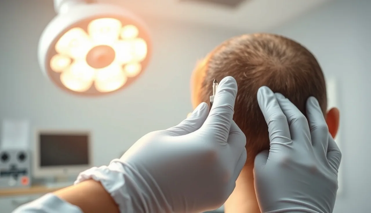
TL;DR: A scalp biopsy removes a small plug of scalp skin (usually 4 mm) and lets a dermatopathologist examine hair follicles under a microscope. It can definitively diagnose androgenetic alopecia, scarring alopecias like lichen planopilaris, alopecia areata, and telogen effluvium when clinical examination alone is inconclusive. Results usually take one to two weeks.
What is a scalp biopsy and why would a dermatologist order one?
A scalp biopsy is a minor in-office procedure where a dermatologist uses a circular punch tool, almost always 4 mm in diameter, to remove a small cylinder of skin from the scalp down to the subcutaneous fat. That tissue goes to a dermatopathologist who slices it horizontally and vertically, stains it, and counts follicles under a microscope. The whole appointment takes under 30 minutes.
Dermatologists order it when they cannot confidently tell two or more types of hair loss apart from a visual exam alone. A receding hairline in a 35-year-old man is usually obvious. But diffuse thinning across the whole scalp in a 42-year-old woman could be androgenetic alopecia, telogen effluvium, early scarring alopecia, or something systemic. Getting that wrong means giving the wrong treatment for months. A biopsy ends the guessing.
The American Academy of Dermatology says dermoscopy and clinical pattern are enough to diagnose androgenetic alopecia in many patients, and that biopsy provides histologic confirmation when the diagnosis is uncertain [1]. That word "uncertain" is doing a lot of work. In one retrospective series of patients referred for hair loss evaluation, clinical diagnosis alone was revised after biopsy in roughly a quarter of cases [2].
Here is the caveat nobody tells you upfront. A biopsy tells you what is happening to the follicle right now. It does not predict how much hair you will lose next year, and it is not a treatment.
How is a scalp biopsy done and does it hurt?
It hurts about as much as a numbing shot at the dentist, then nothing. The dermatologist picks the biopsy site carefully, usually from an area of active hair loss rather than a completely bald patch, because a bald patch has no follicles left to examine. For most diffuse conditions, two punch biopsies come from the same area, one processed horizontally and one vertically, because the two orientations reveal different things about follicle structure and the ratio of terminal to vellus hairs [3].
Locally injected lidocaine numbs the site in about 30 seconds. The punch tool takes one or two seconds. The wound is tiny and usually needs a single suture, or sometimes just a bandage if the site is small. Most people describe the injection as a brief sting and the procedure itself as pressure with no pain.
Healing takes about a week. There is a small scar, but it is usually hidden by surrounding hair. Infection risk is low. If you have a history of keloid scarring, tell your dermatologist before agreeing to a biopsy.
Site selection matters enormously. Biopsying a burnt-out scar with no remaining follicles gives you useless tissue. That is why an experienced dermatologist will often use dermoscopy first to find the inflammatory edge of a scarring condition before picking the biopsy spot [1].
What conditions can a scalp biopsy diagnose?
This is the core question. The table below lists the main diagnoses and their classic biopsy findings.
| Condition | Key biopsy finding |
|---|---|
| Androgenetic alopecia (AGA) | Increased miniaturized follicles, terminal-to-vellus ratio under 4:1, no significant inflammation |
| Telogen effluvium | Elevated telogen count (above 20-25%), normal follicle size, no scarring |
| Alopecia areata | Peribulbar lymphocytic infiltrate ("swarm of bees" pattern), preserved follicle architecture |
| Lichen planopilaris (LPP) | Perifollicular fibrosis, lichenoid lymphocytic infiltrate at isthmus, follicle destruction |
| Discoid lupus erythematosus (DLE) | Follicular plugging, basement membrane thickening, interstitial mucin |
| Central centrifugal cicatricial alopecia (CCCA) | Premature desquamation of inner root sheath, lamellar fibroplasia |
| Folliculitis decalvans | Neutrophilic infiltrate, tufted folliculitis pattern |
| Tinea capitis | Fungal hyphae in hair shaft visible on PAS stain |
The most important split a biopsy makes is between non-scarring alopecias, where follicles are intact and theoretically reversible, and scarring alopecias, where follicle architecture is permanently destroyed. That single distinction changes treatment urgency. Scarring alopecias need aggressive early treatment to stop further loss. Non-scarring alopecias give you more time for a measured approach [4].
Androgenetic alopecia is the most common diagnosis a biopsy confirms. A terminal-to-vellus hair ratio below 4:1 in the frontal scalp is consistent with AGA [3]. If you are already weighing options like finasteride or minoxidil for men, a confirmed AGA diagnosis means those treatments are aimed at the right target. If the biopsy shows LPP instead, finasteride will not touch it and you need anti-inflammatory treatment fast.
What does a scalp biopsy NOT tell you?
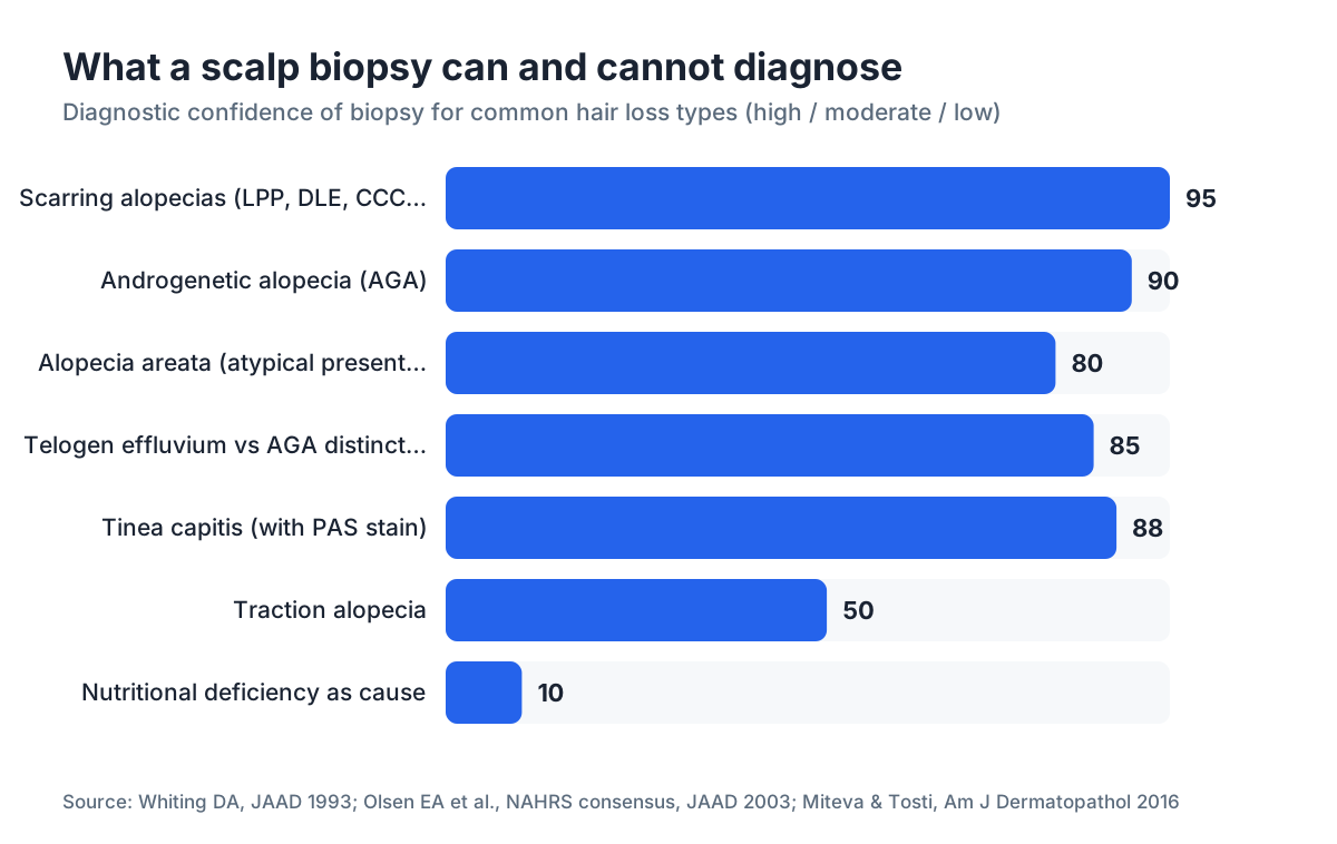
Plenty. A biopsy cannot tell you why you lost hair, only what is happening at the follicle right now. Nutritional deficiencies, thyroid disease, autoimmune antibodies, iron deficiency, and hormonal imbalances get diagnosed through blood tests, not tissue samples. A thorough hair loss workup almost always includes both.
A biopsy also cannot stage the severity of your loss or predict its trajectory. For staging patterns on the scalp, clinicians use the Norwood-Hamilton scale for men and the Ludwig scale for women. Those are clinical assessments, not pathologic ones.
If you have a clear clinical and dermoscopic picture of androgenetic alopecia with a strong family history and the classic pattern of a receding hairline or vertex thinning, most dermatologists will not order a biopsy first. It is a confirmatory and differential tool, not a routine screen.
Results also depend heavily on who reads the slide. Dermatopathology is a subspecialty. A general pathologist may miss subtle early LPP or miscount telogen follicles. Sending samples to a lab with dedicated hair pathology expertise matters, especially for the scarring alopecias.
How accurate is a scalp biopsy at identifying hair loss causes?
Accuracy varies by condition. For androgenetic alopecia with two properly oriented biopsies, the yield is high. A study in the Journal of the American Academy of Dermatology reported that horizontal sectioning correctly identified AGA in over 90 percent of cases when combined with clinical evaluation [3].
For scarring alopecias, biopsy is essentially mandatory because clinical and dermoscopic features overlap heavily between LPP, DLE, CCCA, and other primary cicatricial alopecias. The North American Hair Research Society recommends biopsy for any suspected primary cicatricial alopecia before starting systemic immunosuppression [4].
For alopecia areata, the diagnosis is usually clinical, but in atypical presentations the "swarm of bees" peribulbar infiltrate pattern on histology is distinctive enough to rely on [5].
The biggest accuracy limit is sampling error. Hair loss is often patchy and uneven. A 4 mm punch samples a tiny area. Two biopsies from slightly different zones can give different results. That is exactly why biopsy works best paired with good clinical judgment about where to sample.
Nobody has clean data on the false-negative rate across all conditions. The closest we get is retrospective case series, which carry selection bias because they overrepresent the hard cases that got biopsied in the first place.
When does a dermatologist actually recommend a scalp biopsy?
A biopsy is most likely in four situations. Each one shares a theme: the answer changes what you do next.
The diagnosis is genuinely unclear after examination and dermoscopy. Diffuse hair loss with no obvious pattern and normal bloodwork is the classic scenario.
Scarring alopecia is on the differential. If there is any perifollicular redness, follicular hyperkeratosis, or a symptom of burning and itching at the scalp, a dermatologist needs to rule out a destructive process before it kills more follicles.
A patient is not responding to a treatment that should work. If you have been on adequate-dose minoxidil for a year with no response, the diagnosis might be wrong. A biopsy can confirm or redirect.
There is a medicolegal or insurance need for a definitive histologic diagnosis before approving certain treatments.
For the most straightforward AGA cases, especially younger men with a strong family history and the expected Norwood pattern, a clinical diagnosis is usually enough to start treatment. The American Academy of Dermatology's guidance on androgenetic alopecia treats diagnosis as primarily clinical [1]. Routine biopsies for obvious cases are wasted money and an unnecessary procedure.
Want a fast first read before your appointment? The free AI scan at MyHairline analyzes photos and flags whether your pattern looks like clinical AGA or something worth investigating further. It does not replace a biopsy or a clinical exam. It gives you a starting point.
How much does a scalp biopsy cost and does insurance cover it?
Cash prices in the United States typically run $150 to $600 for the procedure itself, plus $100 to $400 for the pathology reading, depending on the lab and the staining panel [6]. Total out-of-pocket if you pay cash: roughly $250 to $1,000.
Insurance coverage depends on the indication. Most major medical plans, including Medicare, cover diagnostic biopsies when there is a documented clinical reason, such as ruling out a scarring process or an autoimmune condition. Purely cosmetic hair loss evaluations may be denied. Ask your dermatologist to use an appropriate ICD-10 diagnosis code and document medical necessity before the procedure.
Medicare reimburses punch biopsy of skin under CPT code 11104 (first biopsy) and 11105 (each additional biopsy in the same session). The Medicare physician fee schedule lists a national average of roughly $130 to $165 for the procedure component alone, with pathology billed separately [7].
If you are paying cash, ask for the self-pay rate upfront. Many dermatology practices have a different, lower rate for patients who skip insurance.
How long do scalp biopsy results take?
Standard turnaround at most labs is 7 to 14 business days. Rush processing is sometimes available for 3 to 5 days at extra cost.
Complex cases that need immunofluorescence staining, which is used to diagnose lupus-related alopecia, take longer because the tissue requires different processing. Your dermatologist should tell you upfront if they are ordering extra stains.
A report on its own is not the finish line. Histopathology reports for hair loss use specific jargon ("perifollicular fibrosis grade 2," "telogen count 28 percent") that needs interpretation next to your clinical picture. Review results in a follow-up appointment, not a rushed phone call.
What happens after the biopsy confirms a diagnosis?
The treatment path depends entirely on what the biopsy found.
For confirmed androgenetic alopecia, you are looking at FDA-approved options: topical minoxidil, oral minoxidil (off-label), finasteride (for men), or a combination. The evidence for finasteride and minoxidil together is stronger than either alone. For severe cases, a hair transplant may eventually come into the picture.
For telogen effluvium, the treatment is finding and fixing the trigger. Nutritional deficiency, thyroid disorder, crash dieting, major illness, or severe stress are the common ones. The shedding usually resolves within 6 to 12 months once the trigger is gone [8]. Understanding what causes hair loss at a deeper level helps here.
For scarring alopecias like LPP or DLE, treatment is anti-inflammatory (hydroxychloroquine, topical or intralesional corticosteroids, sometimes systemic immunosuppressants). The goal is halting progression, not regrowth, because destroyed follicles cannot regenerate.
For alopecia areata, treatments include intralesional corticosteroid injections, topical immunotherapy, JAK inhibitors (baricitinib and ritlecitinib are FDA-approved for severe alopecia areata as of 2022 and 2023 respectively), and others depending on extent [9].
The result also tells you which supplements or DHT blockers make sense. If the diagnosis is LPP rather than AGA, targeting DHT is pointless. If AGA is confirmed, then saw palmetto, finasteride, and other anti-androgen approaches have a rational basis, though the evidence varies a lot by agent. You can approach hair loss supplements with more confidence once you know what you are actually treating.
Can you distinguish telogen effluvium from AGA with a biopsy?
Yes, and this is one of the most useful things a biopsy does. The two conditions often coexist and can look nearly identical on a visual exam, especially in women with diffuse thinning.
In telogen effluvium, the telogen (resting phase) hair count is elevated, typically above 20 to 25 percent of total follicles in the sample, but follicle caliber stays normal. The miniaturized vellus-like follicles that mark AGA are absent or minimal [3].
In AGA, the terminal-to-vellus ratio drops below 4:1 in affected areas, while the telogen count may be only mildly raised. Both conditions can run together, which shows up as miniaturization plus an elevated telogen count.
The distinction is practical. Chronic telogen effluvium does not respond to finasteride or DHT blockers because androgen sensitivity is not the mechanism. Treating it as AGA burns time and money, and can expose a patient to minoxidil side effects with no realistic payoff.
Are there alternatives to a scalp biopsy for diagnosing hair loss?
Dermoscopy (also called trichoscopy) is the closest non-invasive alternative. A trained dermatologist uses a handheld or video dermoscope to examine follicular openings, hair shaft diameter variation, perifollicular scaling, and vascular patterns. For AGA, dermoscopy shows hair diameter diversity and miniaturization. For LPP, it shows perifollicular scale and absent follicular openings. For alopecia areata, it shows yellow dots and black dots (cadaver hairs) [10].
Dermoscopy is fast, carries no procedural risk, and happens at the first appointment. It has replaced biopsy as the first step in many cases. But it has limits. Early scarring alopecia and AGA can look similar under a dermoscope. A skilled trichoscopist can still miss LPP in its early phase. When dermoscopy is inconclusive and the stakes are high (a suspected scarring process), biopsy is still the right call.
Blood tests catch the systemic causes that neither dermoscopy nor biopsy will find. A typical panel includes TSH, free T4, complete blood count, ferritin, total iron binding capacity, ANA, DHEA-S, free testosterone, and zinc. These get ordered alongside a biopsy, not instead of one.
Photographic monitoring over time, sometimes called global photography or macro photography, tracks progression rather than diagnosing the type. It is a management tool.
If you want a structured way to document your hair density before seeing a specialist, the MyHairline AI scan (/scan) lets you photograph your scalp and get an instant pattern analysis. Doing it before your dermatologist visit means you arrive with baseline photos instead of a vague description of what you saw in the mirror.
AI-assisted trichoscopy tools are being studied in academic settings, but as of 2025, no AI diagnostic tool has FDA clearance for hair loss diagnosis. They are adjuncts, not replacements.
What are the risks and limitations of a scalp biopsy?
The procedural risks are small but real. Infection occurs in less than 1 percent of clean skin biopsies. Scarring is inevitable but usually minor and hidden. Keloid formation is possible in predisposed people. Bleeding is easily controlled with pressure. Rarely, the punch nicks a superficial vessel and causes a small hematoma.
The diagnostic limits matter more for most patients. Sampling error, as covered above, is the main one. A second limit is reader variability between pathologists. A study comparing pathologist readings of the same alopecia slides found real disagreement, particularly for early primary cicatricial alopecias, where the inflammatory infiltrate can be subtle [11].
A biopsy also captures a single moment. Hair loss is dynamic. A biopsy taken right after a major shed from acute telogen effluvium looks different from the same scalp six months later, once it has settled. Timing matters.
And a biopsy cannot confirm a cause that is purely behavioral or environmental, like traction alopecia from tight hairstyles or trichotillomania (compulsive hair pulling). Those need a clinical history, not a histologic report.
Sources
- American Academy of Dermatology
- Mubki T et al., "Evaluation and Diagnosis of the Hair Loss Patient," Journal of the American Academy of Dermatology, 2014
- Whiting DA, "Diagnostic and predictive value of horizontal sections of scalp biopsy specimens in male androgenetic alopecia," Journal of the American Academy of Dermatology, 1993
- Olsen EA et al., North American Hair Research Society consensus on cicatricial alopecia, Journal of the American Academy of Dermatology, 2003
- Alkhalifah A, "Alopecia areata update," Dermatologic Clinics, 2013
- Healthcare Bluebook, Skin Biopsy Cost Estimates
- Centers for Medicare and Medicaid Services, Medicare Physician Fee Schedule (CPT 11104/11105)
- Grover C, Khurana A, "Telogen effluvium," Indian Dermatology Online Journal, 2013
- U.S. Food and Drug Administration, drug approvals
- Rudnicka L et al., "Trichoscopy: A New Method for Diagnosing Hair Loss," Journal of Drugs in Dermatology, 2008
- Miteva M, Tosti A, "Dermatopathology of alopecia," American Journal of Dermatopathology, 2016