

TL;DR: A club-shaped (or club) root is the normal shape of a telogen-phase hair. Finding club roots in shed hairs is the defining sign of telogen effluvium, not scarring or permanent loss. The root is a dry, pale, rounded bulb roughly 0.3 mm across. Seeing it means the follicle cycled early into rest, not that it died. Most cases resolve without treatment once the trigger is removed.
What is a club-shaped hair root?
A club root is the bulb at the base of a hair that shed during the telogen (resting) phase of the hair cycle. Under a basic magnifying glass it looks like a small, dry, white or pale-yellow lump about the size of a grain of sand. The shape is what gives it the name: a rounded knob sitting at the end of an otherwise normal hair shaft.
Every follicle cycles through three phases: anagen (active growth, 2 to 6 years), catagen (transition, 2 to 3 weeks), and telogen (rest, roughly 3 months) [1]. During telogen the follicle keratinizes the root end into that club shape and holds the hair in place until a new anagen hair pushes it out. A healthy scalp sheds 50 to 100 telogen hairs a day with no problem at all [2].
The club root is not a sign of a dead follicle. The follicle is still there, still intact, just resting. That distinction drives everything about your prognosis.
An anagen root looks completely different. It has a soft, translucent, gel-like sheath, is often pigmented at the base, and tapers to a point. Pull an anagen hair out forcibly (or watch what happens in a condition called anagen effluvium) and you see that pointed, sheathed root. The two even feel different. A club hair slides out easily. An anagen hair fights you.
What does a club root tell you about telogen effluvium?
Telogen effluvium (TE) is what happens when an abnormally large share of follicles shift into telogen at the same time, triggered by physiological stress [3]. Instead of the normal 5 to 15 percent of hairs resting at any moment, the proportion can jump to 30 percent or higher.
The shed hairs in telogen effluvium almost always show club roots. That is both the clinical sign and the confirmatory finding. A dermatologist or trichologist doing a gentle hair-pull test collects several hairs and examines the roots. The American Academy of Dermatology describes a positive pull test as extracting more than 6 hairs from a bundle of roughly 60, and in active TE most of those hairs will have club roots [2].
Seeing club roots everywhere in your brush or on your pillow is alarming. It looks like your hair is falling out in clumps. But the message the club root sends is reassuring: the follicle went through a normal keratinization process. It did not get destroyed. Compare that with the roots you see in traction alopecia or chemotherapy-induced anagen effluvium, where the root sheath is stripped and often bloody.
One study published in the Journal of the American Academy of Dermatology found that in acute TE, more than 90 percent of shed hairs showed classic club morphology on trichoscopic examination [4]. That figure is why dermatologists use root morphology as a first checkpoint before ordering blood work.
If you are seeing club roots but the shedding has run past 6 months, that is chronic telogen effluvium (CTE). CTE is a separate, less understood entity. The root morphology is the same, but the persistence signals an ongoing trigger rather than a resolved one.
How is a club root different from a normal shed hair root?
This trips people up because every telogen shed hair has a club root. A "normal" shed hair is a club-root hair. The difference is in the volume of shedding, not the shape of any single root.
Here is the practical breakdown:
| Feature | Normal daily shed | Telogen effluvium shed |
|---|---|---|
| Root shape | Club (rounded, dry, pale) | Club (identical shape) |
| Root color | White to pale yellow | White to pale yellow |
| Hairs per day | 50 to 100 [2] | Often 150 to 400+ |
| Scalp density change | Minimal, none visible | Diffuse thinning, widened part |
| Pull test result | Negative (fewer than 6 hairs) | Positive (more than 6 hairs) [2] |
| Recovery | Not applicable | Usually 3 to 6 months after trigger resolves [3] |
Pick a single hair off your sweater and examine the root. A club shape tells you nothing unusual. What signals a problem is collecting 20 or 30 such hairs in a single shower, or spotting a noticeably thinner crown line when your scalp is wet.
One thing to flag: a broken hair with no root at all is different from a club root. Breakage means shaft damage from heat, bleach, or tight braiding, and has nothing to do with telogen effluvium. If you are only seeing breakage mid-shaft with no bulb, the problem is mechanical or chemical damage to the fiber, not follicle cycling.
What causes the follicle to produce a club root prematurely?
The follicle forms a club root whenever it enters catagen and then telogen, on its normal schedule or early. In telogen effluvium the trigger pushes a large cohort of follicles into that transition ahead of schedule. The common triggers [3][5]:
Postpartum hormonal shift is the most studied. Estrogen that kept follicles locked in anagen during pregnancy drops sharply after delivery, releasing them into telogen all at once. Most postpartum TE peaks 3 to 4 months after birth and resolves by 6 to 12 months [3].
Rapid weight loss and caloric restriction, especially below roughly 1,000 to 1,200 kcal per day, can starve the follicle of the protein and iron it needs to hold anagen [5]. That is one reason crash diets reliably cause shedding 2 to 3 months after the restriction starts.
Fever above 39°C (102°F) sustained for several days, including from COVID-19 infection, is a well-documented precipitant. Post-COVID hair loss was reported in one study as affecting up to 22 percent of hospitalized patients, nearly all through TE with club roots on examination [6].
Iron deficiency (serum ferritin below 30 ng/mL by most trichology guidelines, though some researchers argue the threshold should be 70 ng/mL) is both a trigger and a maintenance factor for chronic TE [5].
Thyroid dysfunction, both hypo- and hyperthyroidism, disrupts the signaling that keeps follicles in anagen. A TSH outside the normal range is one of the first blood tests ordered when TE does not resolve.
Medication-induced TE is common and underreported. Anticoagulants (heparin, warfarin), retinoids, and some antidepressants are listed by the FDA as causing hair loss as a known adverse effect [7]. Even minoxidil side effects can include an early shedding phase called dread shed, a TE-like mechanism as dormant follicles shift to anagen and push out old club hairs.
Surgery under general anesthesia causes TE in a meaningful minority of patients, usually 2 to 3 months post-procedure, because the physiological stress of the operation triggers a synchronized shift to telogen.
Psychological stress is real but harder to pin down. Animal models show cortisol signaling can shorten anagen, and patient histories consistently name major life stress as a trigger, though controlled human data are thin.
How do dermatologists use club roots to diagnose telogen effluvium?
The workup for diffuse shedding starts with a clinical history (timeline, diet changes, illness, medications, recent childbirth) and a scalp exam. The pull test and root morphology examination follow [2].
For the pull test, a clinician grasps about 60 hairs between thumb and forefinger roughly 2 cm from the scalp and applies steady traction toward the tip. More than 6 hairs releasing is a positive result. In active TE the count can hit 15 to 20 or more from a single grab, and nearly all of those hairs show club roots.
Trichoscopy (dermoscopy of the scalp) has largely replaced the need to physically examine individual roots in many dermatology offices. Under dermoscopy the scalp in TE shows increased numbers of short upright regrowing hairs, a sign the follicles are cycling back in, and fewer hair fibers per follicular unit. The absence of miniaturized hairs is what separates TE from androgenetic alopecia (AGA) [4].
A trichogram (plucking a standardized sample of hairs and examining all roots under a microscope) gives a precise anagen-to-telogen ratio. Normal is roughly 85 percent anagen, 15 percent telogen. In active TE that ratio can flip to 50:50 or worse [4].
Blood work usually covers complete blood count, serum ferritin, TSH, free T3/T4, a metabolic panel, and vitamin D. Some clinicians also check zinc, B12, and ANA (antinuclear antibody) if autoimmune causes are suspected. The root morphology findings tell the doctor which tests to order first, since classic club-root TE in a 3-month postpartum window does not need a broad autoimmune panel.
Want a first-pass read before your dermatology appointment? MyHairline's free AI scan (/scan) analyzes scalp photos and shed hair patterns, which helps you frame the conversation with your doctor and track whether shedding volume is changing over time.
Can you tell from a club root alone whether your hair will grow back?
Not fully, but a clean club root is a good sign. It means the follicle finished a normal keratinization at the end of telogen. The papilla, the growth-initiating cluster at the base, was not damaged.
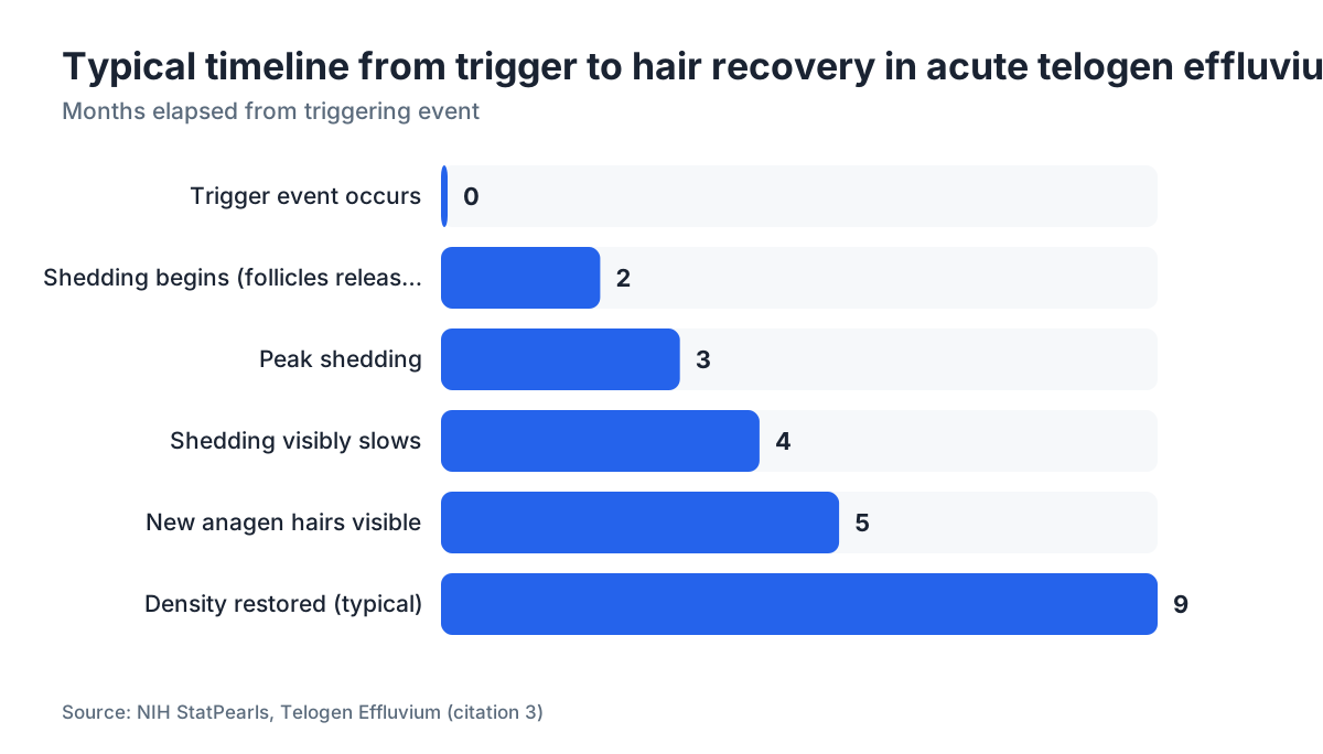
The deciding prognostic factor is whether the underlying trigger has resolved. If the trigger is gone and ferritin, thyroid, and other markers are normal, most people regain their pre-TE density within 6 to 12 months [3]. Regrowth often shows up as short, fine new hairs at the part line 3 to 4 months after the shedding peak.
The reason some people never fully recover usually comes down to one of three things: the trigger was never found and removed, a concurrent androgenetic alopecia was already miniaturizing follicles (so TE made it visible sooner rather than causing it), or the patient has chronic TE with a fluctuating trigger.
One nuance dermatologists flag: if you examine club roots under magnification and the shaft is very thin just above the club, the follicle may have started miniaturizing before the telogen event. A thin club root plus diffuse thinning at the temples and crown should raise suspicion for underlying AGA. That distinction changes the treatment conversation entirely, because AGA needs long-term management with finasteride or minoxidil for men rather than simply correcting a nutritional deficiency.
Age matters too. In women over 40 with diffuse shedding and club roots, CTE and early female pattern hair loss often co-exist, and separating them takes trichoscopy and sometimes a scalp biopsy.
What does a club root look like vs. an anagen root or dystrophic root?
Knowing the visual differences helps you read what you are seeing in your brush or drain.
| Root type | Phase | Appearance | What it means |
|---|---|---|---|
| Club root | Telogen | Dry, rounded, white/pale bulb, no sheath | Normal telogen shed; sign of TE if volume is high |
| Anagen root | Anagen | Soft, gel-like sheath, translucent, often pigmented | Forcible extraction or anagen effluvium (e.g., chemo) |
| Dystrophic/broken root | Damaged anagen | Irregular, frayed, no clear bulb | Active inflammatory or autoimmune alopecia |
| Miniaturized club root | Telogen + miniaturization | Club shape but hair shaft noticeably thinner | AGA follicle entering telogen |
Anagen effluvium, caused by chemotherapy or radiation that poisons rapidly dividing cells, produces anagen roots with that telltale sheath. The shedding is also much faster (days to weeks instead of the 2-to-3-month lag in TE) and often total in the treatment area.
Dystrophic roots look broken or frayed rather than cleanly rounded. They show up in alopecia areata and other inflammatory alopecias. A scalp biopsy is usually needed to confirm those.
For most people reading this: a clean, pale, rounded bulb on your shed hair is a club root, and it points to TE rather than something more destructive.
How long does shedding last and when do club-root hairs stop appearing?
Acute TE follows a predictable arc [3]. The trigger event happens. Two to three months later, shedding peaks. That lag is the follicle's natural telogen duration: the trigger pushed it into telogen at day 0, and telogen runs roughly 100 days before the hair releases. Once the trigger is gone, a new anagen wave starts. You may not see visible improvement in density for another 3 to 6 months, because the new anagen hairs need time to grow long enough to add apparent thickness.
So the full trip from trigger to restored density can run 6 to 12 months even in straightforward cases. This is the part that frustrates people most. Shedding slows well before the scalp looks normal again.
Chronic TE (diffuse shedding lasting more than 6 months) is a different story. CTE can drag on for years if the cause is not found. Commonly missed culprits include subclinical hypothyroidism, celiac disease causing iron and zinc malabsorption, and low-grade chronic psychological stress. In CTE you keep seeing club roots in large numbers with no clear end.
For women, the menstrual cycle and peri-menopause can cause fluctuating TE episodes that feel chronic but are really episodic. Tracking shedding volume with a daily count (collecting all shed hairs from one shower and counting them) beats subjective impression every time.
Children and teenagers get TE too. The same club-root morphology shows up, and the same diagnostic approach applies, though the trigger profile shifts toward infections, nutritional deficiencies from restrictive eating, and rapid growth spurts.
What treatments actually help when telogen effluvium won't resolve?
The honest answer: the most effective treatment is almost always finding and correcting the underlying trigger. No topical or oral medication reliably shortens acute TE while the cause is still active.
For nutritional triggers, iron supplementation (targeting ferritin above 70 ng/mL per some trichology guidelines, though evidence for that specific threshold is observational) and protein intake of at least 0.8 g per kg of body weight per day are the standard dietary moves [5]. There are no large randomized controlled trials on iron and TE specifically. The evidence base is mostly cohort data and expert consensus.
Topical minoxidil for men (and minoxidil for women at 2 percent) is sometimes prescribed in persistent TE or when TE and AGA co-exist. Minoxidil does not treat TE directly. It extends anagen and speeds regrowth of hairs that have already started a new cycle. The FDA has approved 5 percent minoxidil foam for men and 2 percent solution for women for androgenetic alopecia [8]. Using it during TE is off-label but common in practice when recovery is slow.
Oral minoxidil at low doses (0.625 to 2.5 mg daily for women, 2.5 to 5 mg for men) is gaining ground in dermatology practices for diffuse shedding, though the evidence base for TE specifically is still thin.
For TE driven by androgen-sensitive follicles in women, some dermatologists use spironolactone (50 to 200 mg daily), an off-label androgen-blocking approach. For men with concurrent AGA, finasteride and minoxidil together address the AGA component that TE may have unmasked.
Hair loss supplements marketed for TE (biotin, collagen, viviscal-type blends) have weak evidence behind them. A 2019 review in Dermatology and Therapy found biotin deficiency is genuinely rare in healthy adults, and supplementation in non-deficient people does not demonstrably improve hair density [9]. That does not make every supplement useless, but none of them substitute for correcting ferritin, thyroid, or caloric deficits.
Platelet-rich plasma (PRP) injections are used for chronic TE in some practices. The evidence is mixed and the treatment is expensive (typically $500 to $2,500 per session, with multiple sessions needed). What causes hair loss in your specific case should be settled before you spend money on PRP.
Mid-article check-in: if you're trying to sort out whether what you're seeing is TE, AGA, or something else, MyHairline's free AI scan (/scan) analyzes your scalp images and gives you a structured report to take to your dermatologist.
When should you see a doctor instead of waiting it out?
Shedding more than 150 hairs per day for longer than 6 weeks warrants a visit. It is not an emergency, but it is past the normal variation that clears on its own.
See a dermatologist sooner if you notice shedding that started suddenly and worsens each week, visible scalp skin where hair once was, a receding hairline or temple thinning on top of diffuse shedding (which points to AGA co-existing with TE), systemic symptoms like fatigue, cold intolerance, weight change, or menstrual irregularity alongside the shedding, or any scalp redness, scaling, or pain.
The pull test is easy to do at home as a rough check. Grip a bundle of 60 or so hairs, pull firmly toward the ends, and count what comes out. More than 6 club-root hairs is a positive result worth taking to a doctor [2].
Blood work is almost always worth doing at least once. A missed thyroid condition or an iron deficiency left untreated for a year does real cumulative harm beyond the hair, and finding it changes the treatment approach completely.
Pediatric cases and any case where the scalp shows patchy loss rather than diffuse thinning should be seen promptly. Patchy loss suggests alopecia areata or tinea capitis (scalp ringworm), both of which need specific treatment.
Does finding club roots rule out androgenetic alopecia (AGA)?
No, and this is one of the most common diagnostic mistakes people make on their own. Club roots are compatible with both TE and AGA. The two conditions overlap often.
In AGA, follicles undergo progressive miniaturization driven by dihydrotestosterone (DHT) binding to follicle receptors. Each anagen cycle produces a shorter, thinner hair. Those miniaturized hairs eventually shed, and they shed with club roots, same as any telogen hair. The tell is that the club root on an AGA hair sits at the end of a noticeably finer shaft than a healthy telogen hair.
The distinguishing trichoscopic features of AGA are hair shaft diameter variability of more than 20 percent across the affected zone, perifollicular pigmentation (brown halos around follicle openings), and single-hair follicular units in areas where two- and three-hair units should be. These are absent in pure TE [4].
A DHT blocker like finasteride or dutasteride does nothing for TE from iron deficiency or postpartum hormonal change. Fixing ferritin does nothing for genetically miniaturizing follicles. Getting the diagnosis right before choosing a treatment saves time and money.
If you are a man seeing club roots and a receding hairline or thinning crown, odds are high that AGA is the primary driver and TE is secondary. That changes the calculus, because AGA is progressive and needs long-term management, not a 6-month wait for a trigger to clear.
Sources
- National Institutes of Health, StatPearls: Hair Follicle Anatomy
- NIH StatPearls: Telogen Effluvium
- Journal of the American Academy of Dermatology: Trichoscopy in Diffuse Hair Loss
- NIH StatPearls: Nutritional Causes of Hair Loss
- British Journal of Dermatology: Post-COVID-19 Hair Loss
- U.S. Food and Drug Administration, Drug-Induced Hair Loss (MedWatch)
- U.S. Food and Drug Administration, Drugs Database
- Dermatology and Therapy: Biotin Supplementation and Hair Loss (2019 review)
- American Academy of Dermatology, Hair Loss Diagnosis and Treatment