

TL;DR: Telogen effluvium causes diffuse shedding across the whole scalp, not a receding hairline or bald patch. Photos show thinning at the part, a visible scalp under bright light, and handfuls of shed hairs with a white bulb at the root. Most cases resolve within 3 to 6 months once the trigger is gone. It is not permanent in the vast majority of people.
What does telogen effluvium actually look like?
Telogen effluvium looks like the density quietly left the building. No sharp hairline recession. No coin-shaped bald patch. What you see in the mirror, and in photos, is an even thinning that's worst at the crown and along the part. The part gets wider. Bright light reveals scalp that used to stay hidden. Ponytails shrink in circumference. That's the visual signature.
The shed hairs are the other giveaway. Pull one out or pick one off the shower wall and look at the root end under decent light. Telogen hairs have a small, dry, white or pale club-shaped bulb. That bulb means the hair finished its cycle and got pushed out by a follicle that's still alive. Anagen (growing-phase) hairs have a soft, pigmented, sometimes sticky bulb. If you're shedding mostly club hairs, that's strong evidence of telogen effluvium rather than a follicle-destruction process like traction alopecia. [1]
Shedding volume is the number everyone asks about. A normal scalp loses roughly 50 to 100 hairs a day. [2] During a telogen effluvium episode, that can spike to 300 or higher. Some dermatologists use the 100-hairs-a-day mark as a rough cutoff for abnormal shedding, but the honest answer is that counting hairs is hard and the normal range is wider than most sources admit. The sudden change from your own baseline matters more than any fixed number.
What do telogen effluvium photos look like at the part and crown?
The part is the most photographically obvious area. A healthy center part is a thin line with hairs folding naturally to each side. In telogen effluvium, the part looks broad, sometimes three to four times its normal width. Take a top-down photo in direct overhead light and you can trace that widening clearly. The scalp between the remaining hairs reads pink or pale.
The crown tells the same story. From a bird's-eye photo, the hair goes translucent rather than opaque. You can see through the layers to the scalp underneath. That's different from androgenetic alopecia (pattern baldness), where the crown develops a defined bald zone that expands over time. In telogen effluvium the thinning stays even, spread over the whole area, with no single bare spot. [3]
One photography habit beats all the others. Take the same shot weekly under identical light, same time of day, same phone, same angle. A single snapshot fools people constantly because week-to-week variation is huge. Line up six to eight weeks of photos and the real signal appears, recovery or progression.
The front hairline is where people first panic and wrongly decide they're receding. Temporal corners can look slightly thinner during an episode, but the hairline doesn't march backward the way it does in androgenetic alopecia. More on that distinction in our guide to receding hairlines.
How is telogen effluvium different from pattern baldness in photos?
This is the question that causes the most anxiety, and photos alone can't always settle it. But the visual clues are reliable enough to point you in the right direction.
Pattern baldness in men follows the Norwood scale. It progresses from the temples and the crown at the same time, and those two zones can eventually merge into one large bald area. The hair there is genuinely miniaturized, shrunk over years by DHT. Look closely and you'll spot fine, vellus-like strands mixed among the survivors. In photos, the hairline changes shape. The temporal recession goes asymmetric or wedge-shaped. [4]
Telogen effluvium has none of that. The hairline keeps its shape. Remaining hairs aren't miniaturized, there are just fewer of the same caliber. The shedding spreads across the entire scalp, sides and back included, while pattern baldness spares the occipital and temporal fringe. If you're losing hair from the back of your scalp as fast as from the top, that favors telogen effluvium.
Timeline is the other clue photos can capture. Telogen effluvium peaks and then reverses once the trigger clears. Pattern baldness creeps one direction over years. Take dated photos and watch which way the arrow points.
For a fuller look at what could be driving your shedding, our guide to what causes hair loss covers the whole differential.
| Feature | Telogen effluvium | Androgenetic alopecia |
|---|---|---|
| Distribution | Diffuse, whole scalp | Patterned (crown + temples) |
| Hairline | Unchanged shape | Recedes / temples widen |
| Hair shaft caliber | Normal on remaining hairs | Miniaturized hairs visible |
| Timeline | Peaks in weeks, reverses | Slow progression over years |
| Shed hair bulb | White club bulb | Mixed, often no bulb |
| Reversibility | Usually fully reversible | Not without treatment |
What triggers the shedding that shows up in these photos?
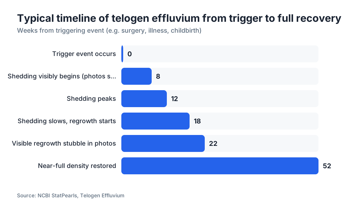
Telogen effluvium happens when a physiological stressor pushes an abnormally large share of follicles out of the growth phase and into the resting (telogen) phase all at once. About two to four months later, those follicles shed their hairs together. That's why photos taken right after a stressful event look fine and photos two months later look alarming. The delay hides the cause. [5]
Common triggers: high fever, surgery, rapid weight loss, giving birth, starting or stopping hormonal contraceptives, severe emotional stress, nutritional deficiencies (iron and ferritin are the best documented), thyroid dysfunction, and certain medications. COVID-19 became one of the most widely reported triggers from 2020 on. A 2021 Lancet Psychiatry study of 236,379 COVID survivors tracked hair loss among the prolonged symptoms following infection. [6]
Chronic telogen effluvium, lasting more than six months, usually has a subtler or ongoing driver: persistently low ferritin, unrelenting psychological stress, or a thyroid condition nobody has caught or that's undertreated. Chronic-case photos show modest but stubborn thinning rather than a dramatic acute shed. People with the chronic form often describe a scalp that has looked thin for a year or more without any single big episode.
If you're trying to separate DHT blocker-relevant pattern loss from a purely effluvium picture, a dermatologist who does trichoscopy (scalp dermoscopy) can tell them apart even when the photographs are ambiguous.
What does recovery from telogen effluvium look like in photos?
Recovery is visible, and it has a look. New hairs come back at the same caliber as before, not fine or miniaturized. They emerge as short hairs that stand up from the scalp, giving the part and crown a fuzzy, bristly texture in photos. Dermatologists call these regrowth hairs. Photograph the part under raking light (a source held low and to the side, not overhead) and you'll see them as a halo of stubble around the part line.
Most acute cases start showing visible regrowth three to six months after the trigger is gone. Full density can take another six to twelve months on top of that, because hair grows roughly half an inch per month. [7] Lose two inches worth of length off the cycle and you're waiting four to five months just to get that length back, even after the follicles restart. This is the part that drives people crazy. Month-four photos can still look thin while recovery is actively underway.
Part width is the single most reliable thing to track. Hold a ruler to the screen. Narrowing means you're recovering. Still widening after six months with the trigger resolved? Book a dermatology visit and rethink the diagnosis. [3]
Some people add minoxidil for men during recovery to try to speed regrowth. The evidence for minoxidil in telogen effluvium is thin next to its evidence base in androgenetic alopecia, but there's no mechanistic reason it would hurt the process.
Can photos alone diagnose telogen effluvium?
No. Photos track change over time, but a diagnosis needs more than a picture. A dermatologist evaluating telogen effluvium typically runs a pull test (grasping 40 to 60 hairs and pulling gently along the shaft, positive being more than 10% releasing as club hairs), and may add a trichogram or trichoscopy. [1]
Blood work carries a lot of weight. Ferritin, complete blood count, thyroid-stimulating hormone, and sometimes zinc and vitamin D make up the standard workup. Ferritin below 30 ng/mL has been linked to hair shedding in several studies, though the exact threshold stays debated in the literature. The American Academy of Dermatology recommends checking ferritin in unexplained diffuse hair loss. [8]
A dated photo series is still genuine clinical data. Diffuse thinning that started two months after a documented trigger (surgery, COVID infection, childbirth) gives a dermatologist something real to work with. Show up to your appointment with photos organized by date. Most dermatologists appreciate it.
If you want a fast first read before you book, the free AI scan at MyHairline analyzes uploaded photos for patterns consistent with diffuse shedding versus androgenetic changes. It's a way to frame better questions for your doctor, not a diagnosis.
What do telogen effluvium photos look like in women versus men?
The general picture is similar in both, but the presentation differs in a few practical ways.
In women, widening at the part is usually the loudest sign. Women's hair tends to be longer, so the diffuse thinning jumps out when hair is wet or pulled back. A ponytail that used to need two loops of a tie now goes three times around. Hairline photos in women with telogen effluvium show a preserved frontal shape, which is the key difference from female pattern hair loss, where a Christmas-tree pattern of widening runs from front to back along the part. [9]
In men, the picture gets muddied by concurrent androgenetic alopecia. A man in his thirties running both conditions at once sees shedding that's worse at the crown and temples, because those follicles are already compromised by DHT sensitivity. The telogen effluvium speeds up the visible progression of the pattern loss. Photos can look more dramatic than either condition would produce alone. That's why men with sudden heavy shedding should get both causes evaluated instead of assuming it's purely one. [3]
For women digging into whether their shedding has a hormonal component, our deeper article on telogen effluvium covers the female-specific triggers, including postpartum and perimenopausal shedding.
What should you look for in the shower drain and on your pillow?
The shower drain is where most people first notice trouble. A normal wash leaves a few hairs against the drain cover. A telogen effluvium shed leaves what looks like a small animal. The hairs run full length or close to it, not broken short, and carry that white club at one end.
Pillow hair is a weaker signal. It depends heavily on how you sleep, how long your hair is, and whether you brush before bed. But if you start waking up to a lot of pillow hair when that never happened before, and it keeps up for weeks, that fits the picture.
Hair on clothing is the subtle one. You glance down at a dark shirt and count dozens of strands. White-tipped hairs support telogen effluvium. Hairs snapped off mid-shaft point to mechanical damage from heat styling or chemical processing rather than anything happening at the follicle.
Perfectly round patches of complete baldness, no hairs at all in a defined circle, are not telogen effluvium. That's alopecia areata, an autoimmune process, and it needs a different workup entirely. [12]
What does a dermatologist's pull test look like on camera?
The pull test is simple and repeatable enough that you can approximate it at home, though a trained eye reads it better. The clinician grasps a bundle of roughly 40 to 60 hairs between thumb and forefinger, applies gentle but firm traction while sliding the fingers from scalp to tip, and counts how many hairs release.
A positive pull test is usually defined as 6 or more hairs releasing out of about 60 pulled (roughly 10%). In active telogen effluvium the count runs much higher. Examine the released hairs under a loupe or microscope: a white club root means telogen, a dark pigmented root means the hair came from the anagen phase and the follicle is being disturbed harder than a pull test should. [1]
On camera, filming a pull test gives you concrete evidence of severity. Video the same section of scalp weekly. Fewer hairs releasing over time is your recovery signal.
A standardized pull test across zones (frontal, parietal, temporal, occipital) also tells you whether the shedding is truly diffuse or concentrated. A positive result across all four zones points hard at telogen effluvium. Positive only at the frontal and parietal zones with a negative occipital pull tilts toward androgenetic alopecia.
Are there any treatments that help telogen effluvium recover faster?
The most effective move is removing or treating the underlying trigger. If it's low ferritin, correcting it with iron has reasonable evidence behind it. A study in the Journal of the American Academy of Dermatology tied non-anemic iron deficiency to chronic diffuse hair loss in premenopausal women. [10] Getting ferritin above 70 ng/mL is a commonly cited clinical target for hair recovery, though that specific number comes from practice rather than a clean randomized trial.
If the trigger is thyroid disease, treating the thyroid usually reverses the shedding over months. Same principle. Fix what caused the problem.
Minoxidil gets recommended sometimes to shorten the regrowth window. The FDA has approved topical minoxidil for androgenetic alopecia (2% for women, 5% for men), not for telogen effluvium, so this is off-label. [11] Some dermatologists reach for it here and some don't. If you try it, know that minoxidil itself causes a temporary shed in the first four to eight weeks as it pushes follicles from telogen into anagen. It looks alarming in photos and is actually a sign it's working.
For the side effect rundown before you start, see our guide on minoxidil side effects. Low-dose oral minoxidil (0.25 to 2.5 mg daily) is increasingly used off-label too; our oral minoxidil guide covers what's known.
Finasteride has no established role in pure telogen effluvium. It blocks DHT-driven miniaturization, which isn't the mechanism here. If you're a man with concurrent pattern loss, finasteride may make sense for the androgenetic component, but it won't speed your TE recovery. Hair loss supplements like biotin get marketed hard, but the evidence for biotin in people without a deficiency is essentially zero. Don't waste money there unless a blood test confirms you're actually low. The finasteride and minoxidil combination is worth knowing about if you're managing overlapping TE and AGA at once.
Our free AI scan can help you track whether your photo-documented shedding is shifting toward a pattern-loss picture that might warrant adding a DHT blocker to the plan.
When do photos suggest you need to see a dermatologist urgently?
Most telogen effluvium is stressful but not an emergency. A few photo findings do earn a faster appointment.
If photos show any completely bald, smooth patches (more than thin areas), that suggests alopecia areata and needs prompt evaluation, because immunosuppressive treatment works best started early. If the scalp skin itself looks off, red, scaly, crusted, or inflamed, that points toward a scarring alopecia or scalp dermatitis, either of which can cause permanent follicle loss if left alone.
If you've tracked photos for six months with a clearly removed trigger and the part is still widening rather than narrowing, revisit the diagnosis. Maybe there's a trigger you haven't found, maybe the chronic TE has a different root like an autoimmune condition, or maybe androgenetic alopecia is progressing under the radar.
Men under 25 with rapid diffuse shedding should get seen. Younger men do get telogen effluvium, but diffuse loss at that age with no obvious trigger warrants ruling out autoimmune causes. Extreme shedding in anyone, where scalp becomes visible within a few weeks rather than months, is unusual enough to warrant an urgent rather than routine visit. The American Academy of Dermatology recommends seeing a board-certified dermatologist for any hair loss that's sudden, patchy, or paired with scalp symptoms. [8]
Sources
- American Academy of Dermatology, Hair Loss Diagnosis and Treatment
- American Academy of Dermatology, Hair Loss Overview
- Hughes EC, Saleh D, Telogen Effluvium, StatPearls (NCBI Bookshelf)
- NIH MedlinePlus, Androgenetic Alopecia
- Hughes EC, Saleh D, Telogen Effluvium, StatPearls (NCBI Bookshelf)
- Taquet M et al., 6-month neurological and psychiatric outcomes in 236,379 survivors of COVID-19, Lancet Psychiatry 2021
- American Academy of Dermatology, Hair Loss Overview
- American Academy of Dermatology, When to See a Dermatologist for Hair Loss
- Olsen EA, Female pattern hair loss, Journal of the American Academy of Dermatology 2001
- Rushton DH, Nutritional factors and hair loss, Clinical and Experimental Dermatology, published on PubMed Central
- U.S. Food and Drug Administration, drug information
- Lepe K, Zito PM, Alopecia Areata, StatPearls (NCBI Bookshelf)